 Open Access Article
Dual heterojunction-based Au@TiO2 photoelectrode exhibiting efficient charge separation for enhanced removal of organic dye under visible light†
Pan
Zhang
a,
Yuzhou
Jin
a,
Mingfang
Li
a,
Xuejiang
Wang
b and
Ya-nan
Zhang
 *a
*a
aSchool of Chemical Science and Engineering, Shanghai Key Lab of Chemical Assessment and Sustainability, Key Laboratory of Yangtze River Water Environment, Tongji University, Shanghai 200092, People's Republic of China. E-mail: yananzhang@tongji.edu.cn
bCollege of Environmental Science and Engineering, Shanghai Institute of Pollution Control and Ecological Security, Tongji University, Shanghai 200092, PR China
First published on 17th March 2023
Abstract
An inventive strategy that involves photo-deposition of Au nanoparticles (NPs) on the flower-like TiO2 nanomicrospheres with co-exposed {001} and {101} facets was reported to construct an advanced visible-light responsive Au@A-FH TiO2/Ti photoelectrode. The plasmonic Au NPs lead to the effective utilization of visible light. The plasmonic Au NPs generate hot e− under visible light and then ransferred to TiO2 through the Au/TiO2 Schottky junction, which finally reach the Ti substrate along the {001}/{101} facet heterojunction (FH). The in situ Kelvin probe force microscopy (KPFM) technology measured the high-resolution contact potential difference (CPD) images between the single Au NP region and the surrounding TiO2 under the visible light, indirectly visualizing the different electron transfer directions between single Au NP and TiO2. Quantitative calculations revealed improved light absorption, charge separation, and surface charge injection efficiency due to the presence of dual heterojunctions (Au/TiO2 Schottky junction and {001}/{101} FH), which together account for high PEC efficiency. The obtained Au@A-FH TiO2/Ti shows high PEC performance under visible light, achieving almost 100% removal of methyl orange (MO) within 2 h. This study provides a set of reliable experimental evidence to verify the electron transfer direction and photoelectrocatalytic mechanism and also offers a new avenue for the design of an efficient TiO2-based visible-light responsive photoelectrode.
Sustainability spotlightWith the intensive development of dyeing, printing, and leather industries, organic dyestuff-based industrial wastewater has caused serious damage to water resources; it is necessary to develop innovative methods to efficiently remove organic dyes in wastewater. The continuing progress of this work is that it proposes an advanced PEC oxidation strategy for enhanced oxidation and complete mineralization of methyl orange under visible light, which depends on the novel Au@TiO2 photoelectrode with dual heterojunctions (Au/TiO2 Schottky junction as well as facet heterojunction). This work is in line with Goal 6 “Clean Water” and 9 “Industrial Innovation”. |
1. Introduction
Against the backdrop of the global ecological water crisis, achieving efficient wastewater treatment and restoration has become an urgent task. As a novel effluent treatment technology, advanced oxidation processes (AOPs) can achieve deep mineralization of pollutants by producing reactive oxygen species (ROS).1,2 Common AOP technologies include ultrasonic oxidation, O3 oxidation, electrocatalysis or photocatalysis; however, they exhibit high energy consumption, easy decomposition (O3 decomposes to O2 in air and water) and poor photoelectric convertibility.3–6 Among AOPs, PEC technology can produce ROS in situ by converting sunlight and applying a bias voltage to massively generate and rapidly separate charges without adding an external catalyst.7,8 The advantages of PEC systems lies in the maximum photocurrent (Jph) can be generated under the minimal bias. This can be achieved by applying photoelectrode materials with the suitable Eg to obtain the highest ηabs (light absorption efficiency), the best ηsep (charge separation efficiency) the maximum ηinj (the efficiency of photogenerated holes injected to the surface reaction).9–11 Therefore, seeking a suitable photoelectrode material is particularly critical.Anatase TiO2 has gained increasing interest because of its low cost, high chemical stability and non-poisonous nature.12,13 Owing to the wide band gap of anatase TiO2 (3.2 eV), it is limited to harvesting UV light, which accounts for a tiny fraction of sunlight. Several methods have been proposed to extend the light absorptivity performance of TiO2, including complication of noble metal nanoparticles (NPs), fabrication of defect states, and sensitization of low-bandgap semiconductors.14–16 Recently, complication of noble metal NPs (e.g., Au, Ag and Pt) has been proposed to extend the light absorption properties of TiO2 owing to their localized surface plasmon resonance (LSPR) effect under certain wavelengths of light excitation.17,18 In particular, Au nanoparticles (NPs) have attracted considerable attention owing to their strong visible light-harvesting ability and high stability. It is claimed that the plasma metal–semiconductor interaction between Au and TiO2 yields the Schottky junction, which facilitates the hot e− transmission from Au NPs to TiO2.19 Further, note that TiO2 still suffers from low quantum inefficiency, the electrons arriving at TiO2 tend to recombinant with each other, leaving a pressing challenge of low ηsep. Constructiing heterogeneous structured photoelectrodes is a method for achieving effective ηsep.20–22 At present, it has been recognized that the crystal facet engineering of TiO2 can also significantly enhance interfacial activity without the introduction of exotic materials, extraordinarily when exposing two facets with multiple band positions matching from one another.23,24 The co-exposed facets form the facet heterojunction (FH), offering intrinsic motivation for the ordered separation of charges. Our recent work found that the anatase TiO2 {001} and {101} facets constitute a typical FH, inducing more photogenerated electron switching from the {001} facet to the {101} facet, thus improving the ηsep.25,26 Therefore, it is promising to load Au NPs onto FH TiO2, the Au NPs composition enables TiO2 to absorb visible light, and the dualle heterojunctions system between Au–TiO2 Schottky junction and {001}–{101} FH achieves rapid separation of photogenerated charges, and the active crystal surface of FH TiO2 promotes the continuous injection of holes, ultimately constructing a high PEC performance photoelectrode.
Herein, we propose to obtain Au@A-FH TiO2/Ti by the photo-deposition of Au NPs on the surface of anatase TiO2 nano–microspheres with {001}/{101} FH. First, the LSPR effect of Au NPs enables the photoelectrode to capture visible light, which significantly extends the visible-light harvesting ability of the photoelectrode. Second, the Au-excited high-energy electrons cross the Au–TiO2 Schottky junction to TiO2, then complete secondary separation along {001}–{101} FH and reach the counter electrode. Finally, the Ti mesh facilitates the fast mass transportation of the solution and effectively increases the area of contact between the contaminant and photoelectrode. The PEC oxidation performance of the photoelectrode was evaluated by eliminating the typical organic dye MO and other non-dye pollutants.
2. Experimental
2.1 Materials
Bisphenol A (≥99.5%), hydrofluoric acid (HF, ≥38.0%), methyl alcohol (CH3OH, >99.9%), nitric acid (HNO3, 65.0–68.0%) and isopropyl alcohol (IPA, >99.0%) were all provided by Sinopharm (Shanghai, China). Absolute ethyl alcohol (CH3CH2OH, >99.9%), anhydrous sodium sulfate (Na2SO4, >99.0%) and anhydrous sodium sulphite (Na2SO3, >99.0%) were obtained from Titan Technologies Inc. (Shanghai, China). Chloroauric acid hydrate (HAuCl4·xH2O, Au: 50%) was bought from Adamas Reagent Co., Ltd. (Shanghai, China). Titanium foil substrate (80 mesh, 0.1 mm in diameter, ≥99.99%) was obtained from Anping Kangwei Wire Mesh Products Co., Ltd. The PTFE hydrothermal kettle was purchased from ShanghaiYanzheng Experimental Instrument Co., Ltd. All reagents were used without further purification.2.2 Synthesis of Au@A-FH TiO2/Ti and Au@A-TiO2/Ti
The titanium mesh substrate needs to be pretreated before participating in the hydrothermal reaction: 3.5 × 5 cm Ti mesh was put into the mixed solution (VH2O![[thin space (1/6-em)]](https://www.rsc.org/images/entities/char_2009.gif) :
:![[thin space (1/6-em)]](https://www.rsc.org/images/entities/char_2009.gif) VHNO3
VHNO3![[thin space (1/6-em)]](https://www.rsc.org/images/entities/char_2009.gif) :
:![[thin space (1/6-em)]](https://www.rsc.org/images/entities/char_2009.gif) VHF = 20
VHF = 20![[thin space (1/6-em)]](https://www.rsc.org/images/entities/char_2009.gif) :
:![[thin space (1/6-em)]](https://www.rsc.org/images/entities/char_2009.gif) 5
5![[thin space (1/6-em)]](https://www.rsc.org/images/entities/char_2009.gif) :
:![[thin space (1/6-em)]](https://www.rsc.org/images/entities/char_2009.gif) 1) for nearly 30 s and then removed. The obtained Ti mesh was immediately placed into DI water and anhydrous ethanol to ultrasonically clean for 5 minutes each and finally reserved it in ethanol solution for further use. 30 mL DI water and 27 μL HF were added into a 100 mL PTFE hydrothermal kettle sequentially, and the solution was stirred well and then put into the pre-processed Ti mesh. The hydrothermal reaction was carried out at 180 °C for 3 h. Similarly, the pre-treated Ti network was placed in 100 mL H2O2 solution and heated at 100 °C for 1 h. The precursors were cleaned with deionized water and reserved as A-FH TiO2/Ti and A-TiO2/Ti.
1) for nearly 30 s and then removed. The obtained Ti mesh was immediately placed into DI water and anhydrous ethanol to ultrasonically clean for 5 minutes each and finally reserved it in ethanol solution for further use. 30 mL DI water and 27 μL HF were added into a 100 mL PTFE hydrothermal kettle sequentially, and the solution was stirred well and then put into the pre-processed Ti mesh. The hydrothermal reaction was carried out at 180 °C for 3 h. Similarly, the pre-treated Ti network was placed in 100 mL H2O2 solution and heated at 100 °C for 1 h. The precursors were cleaned with deionized water and reserved as A-FH TiO2/Ti and A-TiO2/Ti.
The Au NPs were loaded using the photo-deposition method. The obtained A-FH TiO2/Ti photoelectrode was deposited into a mixture of 100 mL H2O, 5 mL CH3OH and 70 μL (or 35 μL and 140 μL) HAuCl4·4H2O (1 wt%), which were irradiated under a xenon lamp (300 W) and stirred for 1 h. The samples were washed with deionized water and dried at 70 °C for 3 h. The photoelectrodes were then calcined at 450 °C for 3 h in the air (the heating rate was 3 °C min−1). The final photoelectrodes are named as 1%Au@A-FH TiO2/Ti (35 μL), 2%Au@A-FH TiO2/Ti (70 μL) and 4%Au@A-FH TiO2/Ti (140 μL). Subsequently, the optimal Au loading was combined with the A-TiO2/Ti to obtain the Au@A-TiO2/Ti contrast photoelectrode.
2.3 Characterization
The topography structure and elemental content of the prepared photoelectrodes were characterized by field emission scanning electron microscopy (SEM, Hitachi-S4800), transmission electron microscopy (TEM, JEM-2100, JEOL) and energy spectrometry (EDS). The elemental valence states on the surface of the photoelectrodes were characterized by X-ray photoelectron spectroscopy (XPS, <10−8 torrent, Al target, 150 W, AXIS Ultra DLD, Shimazu Kratos). XRD diffraction spectroscopy (D8-A25‖ ‖, Bruker) was used to characterize the crystalline shape of the photoelectrodes. The UV-vis absorption range of the photoelectrodes was tested using UV-vis diffuse reflectance spectroscopy (Agilent Carry 5000).2.4 PEC performance test
Photoelectrochemical (PEC) characterization was performed using a standard three-electrode system through the CHI660E electrochemical workstation. The obtained photoelectrodes, platinum sheet and saturated calomel electrode (SCE) were used as the working electrodes, counter electrode and reference electrode, respectively. The electrolyte was 0.1 mol L−1 of Na2SO4 solution. NBeT Solar-500 Xe lamp plus a visible light filter (>420 nm) was used as the light source.2.5 Photoelectrocatalytic experiments
The PEC degradation of MO, phenol and BPA was accomplished by HDY-I potentiostat with the +0.4 V vs. SCE bias voltage and the 50 mL degradation device square double-layered quartz reactor with the external condensate flow, ensuring that the temperature of the system was maintained at room temperature throughout the degradation process. The 45 mL degradation solution contained 5 mg L−1 MO and 0.1 mol L−1 Na2SO4. The initial concentration of phenol and BPA was 1 mg L−1. The light source was a 300 W xenon lamp (PLS-SXE 300) plus a visible light filter (>420 nm) whose output light intensity was 520 mW cm−2. The concentration variations of MO, phenol and BPA during the reaction were determined by applying a UV spectrophotometer (UV-1800), GC (Shimadzu 2014) and HPLC (Agilent 1260). Total organic carbon (TOC) in the post-reaction solution was determined using a total organic carbon analyzer (multi N/C 3100, Analytik Jena, Germany). The scavenging experiment was conducted under the same conditions of PEC degradation, except for adding isopropyl alcohol (IPA) and plenty of nitrogen (N2) as the scavengers of ˙OH and ˙O2−, respectively.2.6 KPFM measurement
The A-FH TiO2/Ti powder distributed in the hydrothermal reaction solution was collected by centrifugal drying. 20 mg A-FH TiO2/Ti powder and 40 mg iodine were dissolved in 100 mL acetone. The FTO substrate and Pt sheet were used separately as the cathode and counter electrode. The uniformly distributed A-FH TiO2/Ti-FTO photoelectrode was energized at an applied potential of +15 V for 15 min. Through the same photoreductive deposition process, the Au@A-FH TiO2/Ti-FTO was obtained and used for KPFM characterization.The spatial morphology and contact potential difference (CPD), i.e., the surface potential, of the Au@A-FH TiO2/Ti-FTO in atmospheric conditions were measured sequentially using KPFM in tap mode. Pt/Ir coated silicon Tips (SCM-PIT-V2) were used as Kelvin tips. UV and visible light incidents perpendicular to the substrate were used to measure the CPD under illumination. The CPD is defined as follows:
| VCPD = (Ws − Wt)/e, |
3. Results and discussion
3.1 Topography characterization and compositional analyses
The TiO2 flower-like microspheres (A-FH TiO2/Ti) were grown in situ on the Ti mesh substrate by applying the hydrothermal method, each with a size of 600–800 nm and consisting of truncated tetra-pyramidal TiO2 nanocrystals (Fig. 1a1 and a2). The relevant morphologies of x%Au@A-FH TiO2/Ti (x = 1, 2, 4) in Fig. 1b1–d2 shows the average size of Au NPs increases gradually with an increased concentration of Au precursor solution (HAuCl4·xH2O) from 1% to 4%. Among them, 2%Au@A-FH TiO2/Ti shows the most uniform distribution in which the Au NPs are evenly coated on the TiO2 microspheres surface (Fig. 1b and S1†), while for 4%Au@A-FH TiO2/Ti (Fig. 1d), the Au NPs tend to agglomerate and obscure TiO2 microspheres. The HRTEM images taken at the 2%Au@A-FH TiO2/Ti (Fig. 1e and f) further confirm the effective loading of Au NPs. The Au particle size distribution, as shown in Fig. S2,† displays that the average size of Au NPs is around 15.7 nm. The HRTEM, as illustrated in Fig. 1g, exhibits that the interplanar crystal spacing is 0.351 nm, corresponding to the {101} facet of anatase TiO2, which can be inferred as the side facet of typical truncated pyramidal.27 The angle between the side facet and the top cross-facet is 122.5°, combined with the model schematic (Fig. S3, ESI†) to determine the top cross-facet is {001} facet.28,29 The exposure percentage of {001} facet is calculated to be approximately 70%, which results in more Au NPs being deposited to the {001} facet.30,31 The well-identified lattice spacings of 0.235 nm on the nanoparticles (Fig. 1h) correspond to the {111} facet of cubic Au.32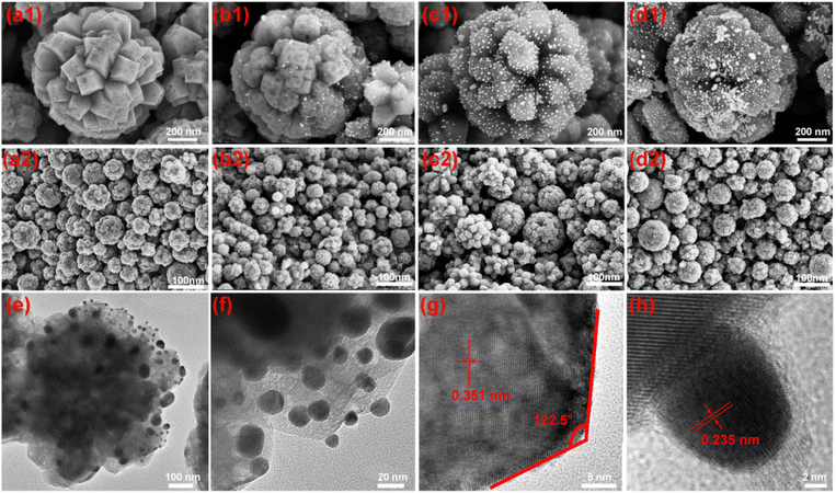 | ||
| Fig. 1 (a1–d2) SEM images of A-FH TiO2/Ti, 1%Au@A-FH TiO2/Ti, 2%Au@A-FH TiO2/Ti and 4%Au@A-FH TiO2/Ti. (e–h) HRTEM images of 2%Au@A-FH TiO2/Ti. | ||
The X-ray diffraction (XRD) pattern (Fig. S4a, ESI†) shows characteristic peaks at 62.7°, 48°, 37.8° and 25.3°, attributing to the (204), (200), (004) and (101) planes of anatase TiO2 (JCPDS no. 21-1272), respectively, indicating that A-FH TiO2 is highly crystalline with anatase structure.33 Compared with the pristine A-FH TiO2, the characteristic crystal phases remain the same after Au loading, except that the intensity of individual peaks is appropriately weakened. The Raman spectra show that the vibrational peaks of all samples belong to the anatase phase of TiO2 (Fig. S4b†).34 Few vibrational peaks belong to Au presumably due to the low content and good dispersion of Au NPs.
X-Ray photoelectron spectroscopy (XPS) was employed to illustrate the elemental composition and chemical states of A-FH TiO2/Ti before and after Au NPs deposition (Fig. 2a); the statistics are listed in Table S2 (ESI†). The characteristic peaks of Ti 2p and O 1s show a positive shift of 0.2 eV and 0.3 eV after Au deposition, respectively (Fig. 2b and c). This suggests the loss of electrons from the Ti and O atoms.35 In addition, the deposition of Au leads to a decrease in the raw area ratio of peak Ti–O–H to peak Ti–O–Ti, which correlates with the tiny quantities of oxygen vacancies (VO), indicating that Au NPs are preferentially deposited at the surface VO sites.36 The Au 4f spectra of Au@A-FH TiO2/Ti shows two significant peaks at 83.0 eV and 86.8 eV, attributing to Au0 4f7/2 and Au0 4f5/2, which demonstrates the free metallic state of Au NPs. Notably, the peak of Au0 4f7/2 (83.0 eV) is lower than that of the bulk Au (∼83.8 eV),37 such a change can be explained by the strong Au/TiO2 interaction, which induces the electron transfer from TiO2 to Au in the dark.
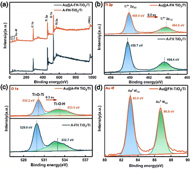 | ||
| Fig. 2 The XPS spectra (a), Ti 2p (b), O 1s (c) and Au 4f (d) spectra of A-FH TiO2/Ti and Au@A-FH TiO2/Ti. | ||
3.2 Photochemical property of photoelectrodes
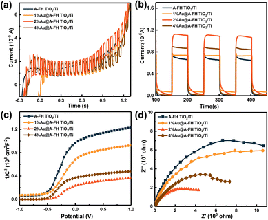
To illustrate the photoelectric conversion capability, the linear sweep voltammograms (LSV) and I–t curves of prepared photoelectrodes under visible-light excitation are shown in Fig. 3a and b, and the relevant data are displayed in Table S1 (ESI†). In all cases, the modified Au@A-FH TiO2/Ti photoelectrode exhibits higher photocurrent density than the unmodified counterparts. Moreover, the photocurrent density of 2%Au@A-FH TiO2/Ti reached the maximum value (1.15 × 10⁻⁵ A cm−2) after three on/off light cycles, which was 1.6, 1.5 and 1.3 times that of A-FH TiO2/Ti, 1%Au@A-FH TiO2/Ti and 4%Au@A-FH TiO2/Ti, respectively. Fig. 3c shows the Mott–Schottky plots produced by the capacitance values. The straight lines of all the photoelectrodes represent positive slopes, indicating typical n-type semiconductor characteristics. Compared to the others, 2%Au@A-FH TiO2/Ti shows the highest carrier concentration (2.67 × 1020 cm−3). As displayed in the typical Nyquist plots (Fig. 3d), the charge transfer impedance of 2%Au@A-FH TiO2/Ti is 5 kΩ, which is significantly smaller than those of 4%Au@A-FH TiO2/Ti (7.5 kΩ), 1%Au@A-FH TiO2/Ti (12 kΩ) and A-FH TiO2/Ti (14 kΩ), suggesting faster charge transfer dynamics. The above results confirm that 2%Au@A-FH TiO2/Ti represents the highest photoelectric response and charge separation efficiency, which is used as the optimal photoelectrode for further study.3.3 The ηabs, ηsep and ηinj of photoelectrodes
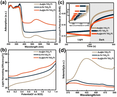
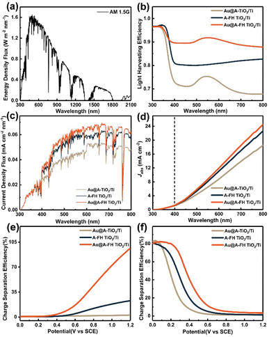
To illustrate the role of Au NPs and facet heterojunction (FH) for PEC performance in detail, A-FH-TiO2 and Au@A-TiO2/Ti (non-specific facet anatase TiO2 loaded with Au NPs in Fig. S5, ESI†) are served as contrast photoelectrodes. The light absorption properties were first investigated by UV-vis diffuse reflectance measurement. As shown in Fig. 4a, in contrast with A-FH TiO2/Ti, the Au NP decoration leads to an extra absorption peak at 550 nm for both Au@A-TiO2/Ti and Au@A-FH TiO2/Ti. This clearly originates from the LSPR effect of Au NPs. Moreover, the absorption intensity of Au@A-FH TiO2/Ti is significantly enhanced once the facet heterojunction between {001} and {101} facets of TiO2 ({001}/{101} FH) is introduced as opposed to Au@A-TiO2/Ti. As depicted in Fig. 4b, the ηabs of the three samples were obtained based on the LSV curves using the following equation:| ηabs = 1 − 10−Jlight | (1) |
Later, the charge separation (ηsep) and injection efficiency (ηinj) of all photoelectrodes are quantified using a hole scavenger of Na2SO3 based on the following equations:
 | (2) |
 | (3) |
 | (4) |
| LHE = 1 − 10−A(λ) | (5) |
The values of ηsep at +0.4 V for Au@A-TiO2/Ti, A-FH-TiO2/Ti and Au@A-FH-TiO2/Ti are 1.1%, 1.3% and 3.9%, respectively (Fig. 5e). The highest ηsep of Au@A-FH-TiO2/Ti is presumably attributed to the synergistic effect of Au/TiO2 Schottky junction and the {001}/{101} FH. Furthermore, Au@A-TiO2/Ti has the highest ηinj of 40.8% at +0.4 V (Fig. 5f), which is much larger than those of Au@A-TiO2/Ti (7.9%) and A-FH TiO2/Ti (20.2%). This suggests the role of oxygen vacancies introduced during the photodeposition of Au NPs, which achieves the effective hole injection between the Au@A-FH-TiO2/Ti and H2O interface. Therefore, the above results demonstrate that the Au NPs and the introduction of FH allow for outstanding PEC catalysis performance.
3.4 In situ KPFM analyses and charge transfer mechanism
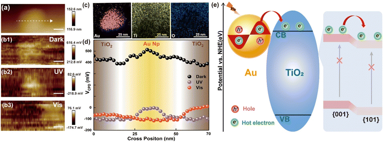
Charge transfer leads to a change in the surface work function in different regions, expressed as a variation in contact potential difference (CPD).40 To further investigate the surface charge transfer process on the surface of of Au@A-FH TiO2/Ti, the in situ KPFM was used to capture the CPD between the single Au NP and TiO2 (Fig. 6a and b) in various environments. Fig. 6a and c separately show the AFM and the TEM-EDS mapping images between single Au NP and TiO2, which agree with the SEM results. Fig. 6b1–b3 show the in situ KPFM potential figures in the dark, UV and visible light, respectively. The brighter color of the KPFM images indicates a higher electron concentration. It can be observed that the surface potential (VCPD) decreases in all cases when the Au@A-FH TiO2/Ti is illuminated. Electron–hole pairs are induced after the Au@A-FH TiO2/Ti is irradiated with a certain wavelength of light, the holes are more inclined to reach the surface, thus reducing the surface electron concentration and leading to lower VCPD. The specific cross-line VCPD values along the dashed arrow (Fig. 6a) between the individual Au NP and TiO2 are summarized in Fig. 6d. Under dark conditions, the VCPD of Au NP is higher than that of the surrounding TiO2, attributing to the higher theoretical work function of Au (5.1 eV) over TiO2 (4.2 eV), which facilitates to form the metal–semiconductor Schottky junction and leads electron to transfer from TiO2 to the surface Au NP. A similar result is obtained under UV illumination, and the VCPD difference in Au/TiO2 interface is more pronounced. This is mainly because TiO2 absorbs the major part of UV light, producing more photogenerated e− to accumulate on Au NP. Notably, in the case of visible light, a situation exactly opposite to the above condition is observed. The VCPD of Au NP is lower than that of the TiO2 nearby. At this time, the high-energy charges transfer from Au NP to TiO2 through Au/TiO2 Schottky junction.By combining the Mott–Schottky curves and the UV-vis diffuse reflectance spectra (Fig. S7, ESI†), the conduction band (ECB) and valence band (EVB) positions of TiO2 were calculated to be −0.75 V and 2.42 eV (vs. NHE),41 respectively. According to the related literature and our previous studies, the energy band arrangement between {001} and {101} facets of TiO2 presents a good step-like matching, which develops {001}/{101} FH.25,42 Therefore, the charge transfer mechanism between Au and TiO2 under visible light is proposed in Fig. 6e. At this time, only Au NP can be excited to generate plasmonic-induced electrons, which further cross the Au/TiO2 Schottky junction and then injected into the TiO2. For the Au NPs deposited on the highly exposed {001} facet, the energetic electrons from the Au LSPR effect fail to reflow owing to the Au–TiO2 Schottky barrier and continue to move from {001} facet to {101} facet along the {001}/{101} FH. The {101} facet acts as a secondary separation intermediate station, which allows electrons to finally arrive at the Ti network substrate. However, for the few Au NPs loaded onto the {101} facet, they are poorly excited to provide enough electrons because of the small exposure ratio and the mutual blockage of {101} facets. Through the synergy of Au/TiO2 Schottky junction and {001}/{101} FH, the plasmonic-induced charges achieve efficient separation and become capable of facilitating efficient PEC reactions.
3.5 Removal of MO under the visible light
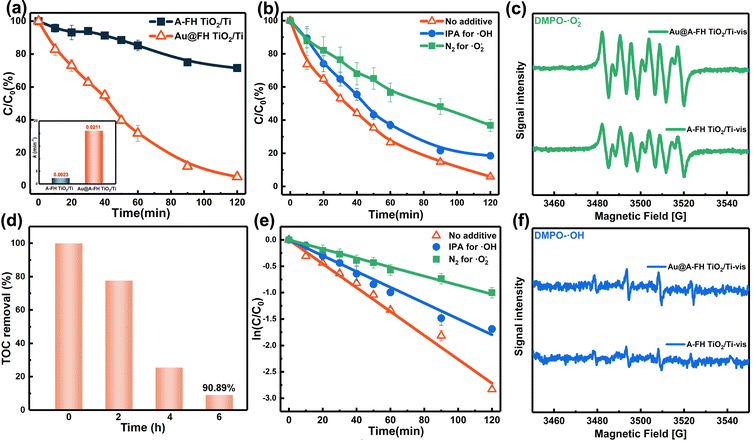
The PEC oxidation ability of Au@A-FH-TiO2/Ti was verified by treating the typical dye contaminant methyl orange (MO) under visible light. As shown in Fig. 7a, the negligible degradation ability of A-FH TiO2/Ti is owing to its wide band gap (3.17 eV), resulting in virtually non-absorption of visible light (Fig. S8, ESI†). As expected, the removal efficiency of Au@A-FH-TiO2/Ti towards MO within 2 h is 94.3% (Fig. 7a) with a rate constant of 0.021 min−1 (the inset in Fig. 7a), which is 3.7 times as high as that obtained over A-FH TiO2/Ti. Similarly, the total organic carbon (TOC) removal efficiency of Au@A-FH-TiO2/Ti for MO reaches 90.89% within 6 h, showing remarkably thorough mineralization ability (Fig. 7d). Afterwards, the main active species in the degradation process under visible light are studied. N2 and isopropanol (IPA) are used as the quenchers of ˙O2− and ˙OH, respectively. Fig. 7b and e indicate that the quenching of ˙O2− (indirectly produced by O2 and e−) significantly inhibits the degradation efficiency of MO from 93.9% to 60.6%, quenching off the ˙OH directly from h+ would slightly reduce the degradation efficiency by 11.5%, which affirms the dominant role of ˙O2− in the degradation process. The corresponding equations are as follows:42| TiO2 + hν ⇌ h+ + e− | (6) |
| h+ + H2O ⇌ ˙OH + H+ | (7) |
| e− + O2 ⇌ O2˙− | (8) |
The electron paramagnetic resonance (EPR) test was used to further verify the generation of ˙O2− and ˙OH. As illustrated in Fig. 7c and f, the ˙O2− and ˙OH signal of Au@A-FH-TiO2/Ti is significantly stronger than that of A-FH TiO2/Ti. Moreover, the signal of ˙O2− is stronger than that of ˙OH. The above results are speculated to be the reason for the high PEC performance.
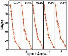
Moreover, five reuse cycles of MO degradation are performed over Au@A-FH-TiO2/Ti, and 91.6% removal of MO was still achieved with no significant changes on the photoelectrode surface (Fig. 8 and S9, ESI†). The XPS spectra in Fig. S10 (ESI†) show that the spectra of Ti 2p, O 1s and Au 4f before and after the PEC reaction are almost identical. These results collectively indicate the good stability and reusability of the prepared photoelectrode. Considering the presence of some colorless organic pollutants in the actual wastewater,43 such as phenol and bisphenol A (BPA), the photoelectrode was also used to handle these pollutants. The results in Fig. S11† suggested that the Au@A-FH TiO2/Ti removed 93.6% of phenol and 95.5% of BPA within 3 h, indicating promising application potential.
4. Conclusion
In conclusion, Au@A-FH-TiO2/Ti photoelectrode was obtained by the photoreduction loading of Au NPs on the surface of flowerlike TiO2 nano–microspheres with {001}/{101} FH. The charge transfer directions in the dark, UV and visible environments were clarified using in situ KPFM tests. Au@A-FH-TiO2/Ti photoelectrode achieved almost 100% removal of MO under visible light in 2 h, and the degradation efficiency was still maintained at 91.6% after five cycles. The LSPR effect of Au NPs broadens the spectral absorption range. The Au/TiO2 Schottky junction and energy-level staggered {001}/{101} FH synergistically facilitate the fast charge separation, thus yielding the highly efficient PEC performance of Au@A-FH-TiO2/Ti. This work provides a reliable and intuitive experimental methodology to verify the charge transfer mechanism, and also provides a novel channel for designing efficient TiO2-based visible light responsive photoelectrodes for the efficient removal of organic dyes and other non-dye pollutants.Author contributions
Pan Zhang: writing-original draft, conceptualization, investigation, formal analysis; Yuzhou Jin: investigation, formal analysis; Mingfang Li: technical assistance, writing-review; Xuejiang Wang: investigation, technical assistance, Ya-nan Zhang: investigation, funding acquisition, writing-review.Conflicts of interest
There are no conflicts to declare.Acknowledgements
This work was supported by National Natural Science Foundation of China (NSFC, No. 22276136, 21876133), Natural Science Foundation of Shanghai (No. 20ZR1462500), the Science & Technology Commission of Shanghai Municipality (No. 14DZ2261100) and the Fundamental Research Funds for the Central Universities (No. 22120210531, 2022-4-YB-1B).Notes and references
- J. Zhang, G. Zhang, H. Lan, J. Qu and H. Liu, Environ. Sci. Technol., 2021, 55, 3296–3304 CrossRef CAS PubMed.
- M. G. Lee, J. W. Yang, H. Park, C. W. Moon, D. M. Andoshe, J. Park, C. K. Moon, T. H. Lee, K. S. Choi, W. S. Cheon, J. J. Kim and H. W. Jang, Nano-Micro Lett., 2022, 14, 48 CrossRef CAS PubMed.
- Y. Yang, J. J. Pignatello, J. Ma and W. A. Mitch, Environ. Sci. Technol., 2014, 48, 2344–2351 CrossRef CAS PubMed.
- D. Yuan, M. Sun, S. Tang, Y. Zhang, Z. Wang, J. Qi, Y. Rao and Q. Zhang, Chin. Chem. Lett., 2020, 31, 547–550 CrossRef CAS.
- A. K. Biń and S. Sobera-Madej, Ozone: Sci. Eng., 2012, 34, 136–139 CrossRef.
- C. A. Martínez-Huitle and M. Panizza, Curr. Opin. Electrochem., 2018, 11, 62–71 CrossRef.
- Q. Ma, R. Song, F. Ren, H. Wang, W. Gao, Z. Li and C. Li, Appl. Catal., B, 2022, 309, 121292 CrossRef CAS.
- L. Liu, P. Li, T. Wang, H. Hu, H. Jiang, H. Liu and J. Ye, Chem. Commun., 2015, 51, 2173–2176 RSC.
- T. Zhou, S. Chen, L. Li, J. Wang, Y. Zhang, J. Li, J. Bai, L. Xia, Q. Xu, M. Rahim and B. Zhou, Appl. Catal., B, 2020, 269, 118776 CrossRef CAS.
- F. Wu, Y. Yu, H. Yang, L. N. German, Z. Li, J. Chen, W. Yang, L. Huang, W. Shi, L. Wang and X. Wang, Adv. Mater., 2017, 29, 1701432 CrossRef PubMed.
- R. Song, H. Chi, Q. Ma, D. Li, X. Wang, W. Gao, H. Wang, X. Wang, Z. Li and C. Li, J. Am. Chem. Soc., 2021, 143, 13664–13674 CrossRef CAS PubMed.
- Q. Niu, X. Gu, L. Li, Y.-n. Zhang and G. Zhao, Appl. Catal., B, 2020, 261, 118229 CrossRef CAS.
- P. Zhang, X. Gu, N. Qin, Y. Hu, X. Wang and Y. N. Zhang, J. Hazard. Mater., 2023, 441, 129896 CrossRef CAS PubMed.
- X. Zheng, H. Guo, Y. Xu, J. Zhang and L. Wang, J. Mater. Chem. C, 2020, 8, 13836–13842 RSC.
- M. Y. Ye, Z. H. Zhao, Z. F. Hu, L. Q. Liu, H. M. Ji, Z. R. Shen and T. Y. Ma, Angew. Chem., Int. Ed. Engl., 2017, 56, 8407–8411 CrossRef CAS PubMed.
- H. Li and L. Zhang, Nanoscale, 2014, 6, 7805–7810 RSC.
- C. Boerigter, R. Campana, M. Morabito and S. Linic, Nat. Commun., 2016, 7, 10545 CrossRef CAS PubMed.
- S. T. Kochuveedu, Y. H. Jang and D. H. Kim, Chem. Soc. Rev., 2013, 42, 8467–8493 RSC.
- Z. Bian, T. Tachikawa, P. Zhang, M. Fujitsuka and T. Majima, J. Am. Chem. Soc., 2014, 136, 458–465 CrossRef CAS PubMed.
- A. Kumar, M. Khan, J. He and I. M. C. Lo, Water Res., 2020, 170, 115356 CrossRef CAS PubMed.
- H. Zhao, X. Liu, Y. Dong, Y. Xia, H. Wang and X. Zhu, ACS Appl. Mater. Interfaces, 2020, 12, 31532–31541 CrossRef CAS PubMed.
- D. Gogoi, A. K. Shah, P. Rambabu, M. Qureshi, A. K. Golder and N. R. Peela, ACS Appl. Mater. Interfaces, 2021, 13, 45475–45487 CrossRef CAS PubMed.
- Y. Hu, Y. Jin, P. Zhang, Y.-n. Zhang and G. Zhao, Appl. Catal., B, 2023, 322, 122102 CrossRef CAS.
- J. Yu, J. Low, W. Xiao, P. Zhou and M. Jaroniec, J. Am. Chem. Soc., 2014, 136, 8839–8842 CrossRef CAS PubMed.
- S. Han, Q. Niu, N. Qin, X. Gu, Y. N. Zhang and G. Zhao, Chem. Commun., 2020, 56, 1337–1340 RSC.
- X. Gu, N. Qin, G. Wei, Y. Hu, Y. N. Zhang and G. Zhao, Chemosphere, 2021, 263, 128257 CrossRef CAS PubMed.
- C. P. Sajan, S. Wageh, A. A. Al-Ghamdi, J. Yu and S. Cao, Nano Res., 2015, 9, 3–27 CrossRef.
- J. Yu, G. Dai, Q. Xiang and M. Jaroniec, J. Mater. Chem., 2011, 21, 1049–1057 RSC.
- X. Liu, G. Dong, S. Li, G. Lu and Y. Bi, J. Am. Chem. Soc., 2016, 138, 2917–2920 CrossRef CAS PubMed.
- H. Zhang, X. Liu, Y. Wang, P. Liu, W. Cai, G. Zhu, H. Yang and H. Zhao, J. Mater. Chem. A, 2013, 1, 2646–2652 RSC.
- M.-Y. Xing, B.-X. Yang, H. Yu, B.-Z. Tian, S. Bagwasi, J.-L. Zhang and X.-Q. Gong, J. Phys. Chem. Lett., 2013, 4, 3910–3917 CrossRef CAS.
- J. Yu, G. Dai, Q. Xiang and M. Jaroniec, J. Mater. Chem., 2011, 21, 1049–1057 RSC.
- Y. Lu, X. Liu, H. Liu, Y. Wang, P. Liu, X. Zhu, Y. Zhang, H. Zhang, G. Wang, Y. Lin, H. Diao and H. Zhao, Small Struct., 2020, 1, 202000025 Search PubMed.
- L. K. Preethi, T. Mathews, M. Nand, S. N. Jha, C. S. Gopinath and S. Dash, Appl. Catal., B, 2017, 218, 9–19 CrossRef CAS.
- J. Qi, X. Yang, P. Y. Pan, T. Huang, X. Yang, C. C. Wang and W. Liu, Environ. Sci. Technol., 2022, 56, 5200–5212 CrossRef CAS PubMed.
- R. Siavash Moakhar, G. K. L. Goh, A. Dolati and M. Ghorbani, Appl. Catal., B, 2017, 201, 411–418 CrossRef CAS.
- D. Ding, K. Liu, S. He, C. Gao and Y. Yin, Nano Lett., 2014, 14, 6731–6736 CrossRef CAS PubMed.
- G. Yang, Y. Li, H. Pang, K. Chang and J. Ye, Adv. Funct. Mater., 2019, 29, 1904622 CrossRef CAS.
- H. Li, S. Wang, M. Wang, Y. Gao, J. Tang, S. Zhao, H. Chi, P. Zhang, J. Qu, F. Fan and C. Li, Angew. Chem., Int. Ed. Engl., 2022, 61, e202204272 CAS.
- R. Chen, Z. Ren, Y. Liang, G. Zhang, T. Dittrich, R. Liu, Y. Liu, Y. Zhao, S. Pang, H. An, C. Ni, P. Zhou, K. Han, F. Fan and C. Li, Nature, 2022, 610, 296–301 CrossRef CAS PubMed.
- T. Zhou, S. Chen, L. Li, J. Wang, Y. Zhang, J. Li, J. Bai, L. Xia, Q. Xu, M. Rahim and B. Zhou, Appl. Catal., B, 2020, 269, 118776 CrossRef CAS.
- L. Hu, C. C. Fong, X. Zhang, L. L. Chan, P. K. Lam, P. K. Chu, K. Y. Wong and M. Yang, Environ. Sci. Technol., 2016, 50, 4430–4438 CrossRef CAS PubMed.
- S. Garcia-Segura and E. Brillas, J. Photochem. Photobiol., C, 2017, 31, 1–35 CrossRef CAS.
Footnote |
| † Electronic supplementary information (ESI) available. See DOI: https://doi.org/10.1039/d3su00006k |
| This journal is © The Royal Society of Chemistry 2023 |

