 Open Access Article
Creative Commons Attribution 3.0 Unported Licence
Identification and functional characterization of novel plant UDP-glycosyltransferase (LbUGT72B10) for the bioremediation of 3,4-dichloroaniline
Valeria
Della Gala
 and
Ditte Hededam
Welner
 *
*
The Novo Nordisk Foundation Center for Biosustainability, Technical University of Denmark, Kemitorvet 220, DK-2800 Kgs. Lyngby, Denmark. E-mail: diwel@biosustain.dtu.dk; Tel: +45 93513498
First published on 25th September 2023
Abstract
Herbicides are chemicals used to manipulate or control the growth of undesired plants and thereby protect crops. However, they and their degradation products can persist and accumulate in the environment, leading to contamination of soil and water systems and biodiversity loss. Interestingly, through the action of UDP-glycosyltransferases (UGTs), higher plants can glycosylate these xenobiotics, increasing their solubility and alleviating their toxicity. Here, seven plant UGTs belonging to family 72 of the UGT nomenclature were identified to N-glycosylate 3,4-dichloroaniline (3,4-DCA), which is a degradation product of commercially significant herbicides like Diuron, Linuron and Propanil. Although chlorinated chemicals are well-known UGT substrates, only one UGT with activity on 3,4-DCA (AtUGT72B1 from Arabidopsis thaliana) has been fully biochemically characterized. In this study, biochemical analysis revealed that six of the seven identified UGTs are capable of full conversion of 3,4-DCA to its N-glucoside. The most efficient enzyme was found to be LbUGT72B10 from Lycium barbarum (kcat = 11.2 s−1, KM = 51.2 μM). Consequently, transgenic expression of LbUGT72B10 could potentially play a role in the future in the mitigation of 3,4-DCA toxicity, preventing its accumulation in living systems and reducing contamination of waterways and soil.
Sustainability spotlightThe persistent environmental contaminant 3,4-dichloroaniline (3,4-DCA) results from the degradation of herbicides and industrial pollutants. UDP-glycosyltransferases (UGTs) can facilitate the bioremediation of 3,4-DCA through glycosylation, promoting its biodegradation in the environment and lowering human exposure to the chemical. This study reports the identification and functional characterization of LbUGT72B10, a novel N-glycosyltransferase active on 3,4-DCA. Although further investigations are required to fully understand the actual bioremediation mechanism, UGT-mediated glycosylation can help mitigate the health risks associated with the pollutant and improve its biodegradation from water and soil sources, resulting in better water quality and reduced biodiversity loss. Therefore, this study addresses various sustainability challenges aligned with the UN Sustainable Development Goals, including SDG 3, 6, 14, and 15, related to improving human well-being, clean water, preserving life below water, and promoting life on land. |
1. Introduction
Chlorinated xenobiotics are a group of artificially produced chemicals characterized by a cyclic structure and a variable number of chlorine atoms.1 They are used as crop-protecting agents against weeds and pests or in the manufacturing of pesticides, and they also play a role in the pulp and paper industry.2,3 They are classified as Persistent Organic Pollutants (POPs) and, due to their lipophilic nature and persistence, they possess high bioaccumulation potential.4–6 These pollutants can enter the ecosystem via a variety of waste streams, but primarily through pesticide application, posing a threat to biodiversity.7 Due to their high hydrophobicity, they accumulate in the fat tissues of animals and in different structures of plants.8,9 Additionally, despite being formulated to target specific categories of weeds, these compounds can be non-selective and cause harm to most plants they come in contact with.10,11 In animals including humans, long-term exposure to these chemicals has been shown to cause neurotoxic effects, liver and kidney damage and possibly cancer.12 Chlorinated anilines are a major class of xenobiotics in the environment.13 They are mainly released in the environment as biodegradation products of economically significant herbicides such as Propanil, Diuron and Linuron.14 Particularly, dichloroanilines, such as 3,4-dichloroaniline (3,4-DCA), persist in the environment for a long time as insoluble residues in soil and plant systems, and are detected more frequently compared to their parent compounds.15–17 3,4-DCA has been detected in surface waters of the European Ebro river basin, with average concentrations ranging from 25 ng L−1 to 765 ng L−1.18 It has also been found in effluents from dye-manufacturing plants and water field cultures in other parts of the world.19,20 Notably, high concentrations of 3,4-DCA have been recorded in water samples, reaching up to 567 μg L−1, and in soil samples from Brazilian rice fields, with levels reaching 119 mg kg−1.21 3,4-DCA is toxic to aquatic organisms and has been shown to induce genotoxic effects, reproductive toxicity and histopathological alteration in mice, being in this way potentially harmful to humans.22 It has also been shown to be more toxic than its parent chemicals.23 Once introduced into the environment, 3,4-DCA is rapidly absorbed by the roots and translocated to the leaves and other parts of the plant.24Interestingly, plants can regulate the biological activity of pollutants, pesticides, and their metabolites by converting them into less toxic compounds through a four-phase detoxification system: first, xenobiotics are metabolically activated by “phase 1” enzymes, which then facilitates their subsequent bioconjugation with polar natural products in “phase 2” metabolism.25 In crops and weeds, the most observed “phase 2” reaction is glycosylation, which is a biochemical modification catalyzed by a family of enzymes known as UDP-glycosyltransferases (UGTs).26 Plant UGTs catalyze the transfer of active sugar groups from nucleotide sugars, usually from uridine diphosphate-glucose (UDP-Glc) to small molecules, such as phytohormones and secondary metabolites, as well as xenobiotics (Fig. 1).27–30 In “phase 3”, the glycosides can either be imported into the vacuoles or they can go through additional conjugation with malonyl-CoA, which is a frequent reaction in higher plants that results in the formation of non-phytotoxic secondary conjugates.31,32 A study has shown that the root culture of Arabidopsis thaliana can quickly conjugate 3,4-dichloroaniline to 3,4-dichloroaniline-N-glucoside, which is then transported outside the roots.33 The vacuolar storage and the release into the rhizosphere reduce the phytotoxicity of the xenobiotic and prevent yield losses. Therefore, by increasing the water solubility, glycosylation of xenobiotics can help prevent bioaccumulation.32 Interestingly, the model plant Arabidopsis thaliana contains more than 100 UGTs, which are dedicated to small molecule conjugation, and 44 of them catalyze the O-glycosylation of chlorinated chemicals.31 Multiple studies have confirmed that many UGTs can glycosylate them as well as their natural acceptor.34–37 Nonetheless, only a limited number of UGTs, specifically 10, have been discovered to have low N-glycosylation activity towards 3,4-DCA.13,33,35,36,38 Out of these N-glycosyltransferases (N-GTs), AtUGT72B1 was the only enzyme found to have appreciable N-glycosylation activity, and it was also the only one that has been biochemically characterized with this substrate.31 In the present work, we selected seven candidates from our UGT collection for 3,4-DCA glycosylation, based on sequence identity (>60%) with AtUGT72B1. Of these seven candidates, which belong to the UGT72 family, six showed full conversion of 3,4-DCA to its N-glucoside, and LbUGT72B10 was identified as the most efficient enzyme and biochemically and kinetically characterized. Our in vitro results show that LbUGT72B10 is an efficient biocatalyst for the conversion of the xenobiotic to 3,4-DCA-N-glucoside (Fig. 1). These results highlight the potential value of LbUGT72B10 in bioremediation strategies aimed at removing the pollutant from the environment by utilizing transgenic technology to manipulate xenobiotic metabolism in plants. However, additional in vivo investigations are required to validate and clarify its effectiveness as a potential bioremediation strategy in order to enhance agricultural sustainability.
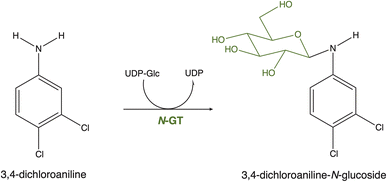 | ||
| Fig. 1 Glycosylation reaction of 3,4-DCA to 3,4-DCA-N-glucoside catalyzed by a N-glycosyltransferase (N-GT). | ||
2. Materials and methods
2.1 Reagents and chemicals
Buffers, standard reagents, and 3,4-dichloroaniline (≥98%) were purchased from Sigma-Aldrich.2.2 Sequence selection
To identify the closest homologs to the known 3,4-DCA-glycosylator AtUGT72B1 (Uniprot Accession: Q9M156, GenBank Accession: AF360262.1) available in our laboratory, the sequence of AtUGT72B1 was used as a query for the NCBI Basic Local Alignment Search Tool (BLASTp) search against our in-house UGT library sequences.39 Our in-house UGT library is a random selection of UGTs from phylogenetic group E and contains 45 sequences from various plant species, which are readily available from prior studies within our group, as plasmids suitable for the expression of soluble recombinant proteins. The candidate sequences were selected based on their >60% sequence identity with the query sequence. Multiple sequence alignment (MSA) was generated using the Clustal Omega (https://www.ebi.ac.uk/Tools/msa/clustalo/) and visualized with Jalview software (http://www.jalview.org/).2.3 Cloning, expression and purification of recombinant UGTs
The full-length coding sequence of the UGTs was synthesized and cloned into the pET28a(+) vector by Genscript (USA), using the NcoI and XhoI restriction sites. The protein coding sequences included an N-terminal sequence consisting of a 6xHis-tag. E. coli BL21 Star™ (DE3) cells (Thermo Fisher Scientific) were transformed with the plasmids and the precultures were grown overnight at 37 °C in LB medium supplemented with 50 μg mL−1 kanamycin. Main cultures were prepared by adding 5 mL overnight culture to 500 mL of 2xYT culture with 50 μg mL−1 kanamycin and grown at 37 °C and 220 rpm until OD600 = 0.5–0.7. The cultures were induced with 0.5 mM IPTG and grown for about 22 hours at 20 °C. The cells were harvested by centrifugation at 4500 × g at 4 °C and resuspended in a buffer containing 50 mM HEPES, 300 mM NaCl, pH 7.5, with 20 mM imidazole supplemented with 0.4 mg DNaseI. The resuspended cells were subjected to cell lysis using an Avestin Emulsiflex C5 homogenizer (ATA Scientific Pty Ltd., Canada), and the soluble cell lysate was recovered by centrifugation at 14![[thin space (1/6-em)]](https://www.rsc.org/images/entities/char_2009.gif) 500 × g at 4 °C for 50 min. The supernatant containing the recombinant proteins was filtered with a 0.22 μM filter and purified by nickel-affinity chromatography (1 mL prepacked HisTrap™ FF columns, GE Healthcare, Sweden) on an ÄKTA pure (GE Healthcare, Sweden). Prior to sample loading, the column was washed and equilibrated with the same buffer (50 mM HEPES, 300 mM NaCl, pH 7.5, with 20 mM imidazole). Following protein binding, the column was washed with a buffer containing 50 mM HEPES, 300 mM NaCl, pH 7.5, with 500 mM imidazole. Elution of the proteins was achieved using a combination of isocratic and gradient elution with an imidazole concentration increasing from 20 to 500 mM. The elution peak fractions were pooled together and concentrated using centrifugal filters (MW cut-off 10–30 kDa), and buffer exchanged to a buffer containing 25 mM HEPES, 150 mM NaCl, pH 7, with 1 mM DTT. The total concentration of partially purified proteins was measured by spectrophotometric measurements at 280 nm in a NanoDrop Instrument (Thermo Fisher Scientific). The sample purity was assessed using SDS-PAGE.
500 × g at 4 °C for 50 min. The supernatant containing the recombinant proteins was filtered with a 0.22 μM filter and purified by nickel-affinity chromatography (1 mL prepacked HisTrap™ FF columns, GE Healthcare, Sweden) on an ÄKTA pure (GE Healthcare, Sweden). Prior to sample loading, the column was washed and equilibrated with the same buffer (50 mM HEPES, 300 mM NaCl, pH 7.5, with 20 mM imidazole). Following protein binding, the column was washed with a buffer containing 50 mM HEPES, 300 mM NaCl, pH 7.5, with 500 mM imidazole. Elution of the proteins was achieved using a combination of isocratic and gradient elution with an imidazole concentration increasing from 20 to 500 mM. The elution peak fractions were pooled together and concentrated using centrifugal filters (MW cut-off 10–30 kDa), and buffer exchanged to a buffer containing 25 mM HEPES, 150 mM NaCl, pH 7, with 1 mM DTT. The total concentration of partially purified proteins was measured by spectrophotometric measurements at 280 nm in a NanoDrop Instrument (Thermo Fisher Scientific). The sample purity was assessed using SDS-PAGE.
2.4 Glycosyltransferase activity assay toward 3,4-dichloroaniline
Partially purified protein samples were used in 30 μL of total reaction volume to perform in vitro glycosylation reactions. The reaction mixture contained 50 μg mL−1 of enzymes, 500 μM 3,4-DCA, 2 mM UDP-Glc in a 50 mM phosphate-citrate buffer, pH 7. The control was made with the reaction mixture without the enzyme. The reaction was incubated at 20 °C and quenched at various time intervals (2 min, 5 min and 10 min) by adding 1 volume of ice-cold acetonitrile. Data were analyzed as mean ± S.D. of two independent experiments.2.5 Biochemical characterization of LbUGT72B10
For the identification of the pH optimum of LbUGT72B10, its activity was investigated in 100 mM Tris–BisTris buffer, pH 5–10, with 2 μg mL−1 enzyme, 50 μM 3,4-DCA and 200 μM UDP-Glc. 10 μL of reaction were quenched with 240 μL 0.1% acetic acid at 0, 2, 4, 6, 8 and 10 minutes.For the identification of the temperature optimum, 2 μg mL−1 enzyme activity towards 50 μM 3,4-DCA were assayed in 100 mM Tris–BisTris buffer, pH 7.75, with 200 μM UDP-Glc. The reactions were carried out at 30–54 °C in a thermocycler and were stopped by the addition of 240 μL acetic acid (0.1%) to 10 μL of the reaction mix after 5, 15, 60 and 180 min. In both cases, data were analyzed as mean ± S.D. of two independent experiments.
For the identification of the melting temperature, differential scanning fluorimetry (DSF) was performed using the Protein Thermal Shift Dye Kit (Thermo Fisher Scientific) and a qPCR QuantStudio5 machine. Dye solution (1000×) was diluted to 2× in 100 mM citrate-phosphate buffer, pH 7. 10 μL of diluted dye solution was mixed with 10 μL of 0.8 mg mL−1LbUGT72B10 in 2× of the same buffer and pipetted into a qPCR 96-wells plate. The plate was centrifuged for 30 s at 1000 rpm and transferred to the qPCR machine. After 2 min incubation at 25 °C, the temperature increased gradually to 99 °C, with a final incubation of 2 min at 99 °C. Measurements were carried out in triplicate. Raw data were analyzed as mean ± S.D. of three independent experiments with Protein Thermal Shift™ Software v1.x.
2.6 Kinetic characterization of LbUGT72B10
For the kinetic characterization, based on the optimized reaction conditions, the activity of 10 ng LbUGT72B10 was assayed towards 5–500 μM 3,4-DCA in 100 mM Tris–BisTris buffer, pH 7.75 at 30 °C. The measurements were carried out under saturating UDP-Glc conditions (1 mM). The reaction was initiated by adding the acceptor substrate and quenched by adding 240 μL acetic acid (0.1%) to 10 μL of the reaction mix after 3 min. The μM of product formed was obtained from the peak area and the corresponding substrate concentration. Data were analyzed as mean ± S.D. of three independent experiments. KM and kcat values were determined by fitting the initial velocity data using the Michaelis–Menten model and the drc package in Rstudio (Rstudio version 2022.12.0 + 353, RStudio, Inc.).2.7 HPLC analysis
In all enzymatic assays, the reaction mixture was analyzed by reverse-phase HPLC using an Ultimate 3000 Series apparatus (Thermo Fisher Scientific) and a kinetex 2.6 μm C18 100 Å 100 × 4.6 mm analytical column (Phenomenex). Milli-Q water containing 0.1% formic acid and acetonitrile were used as mobile phases A and B, respectively. A combination of isocratic, immediate ramp, and gradients at a flow rate of 1 mL min−1 were applied for the separation of 3,4-DCA and 3,4-DCA-N-glucoside: 0–0.5 min, 2% B; 0.5–1.5 min, 35% B; 1.5–3 min, 35–80% B; 3–4.2 min, 98% B; 4.2–5 min, 2% B. The analytes were detected at 260 nm. The HPLC data was monitored and quantified via the Chromeleon software (Thermo Fisher Scientific).3. Results
3.1 Candidate gene selection
To identify additional bioremediation candidates potentially capable of N-glycosylating 3,4-DCA, we selected seven UGT sequences (Table 1) from our “local” enzyme collection with >60% sequence identity to AtUGT72B1 (Uniprot Accession: Q9M156, GenBank Accession: AF360262.1) using NCBI BLASTp search.39 The multiple sequence alignment (MSA) of the selected UGTs and AtUGT72B1 is shown in Fig. 2. All the selected UGTs belong to the UGT72 family, which has been shown to be active with flavonoids, monolignols, chlorophenols, chloroanilines and their precursors or derivatives as substrates.40 The selected UGTs contain the conserved PSPG motif in their C-terminal domain, which is responsible for recognizing the nucleotide-activated sugar (UDP-Glc).41,42 Furthermore, the selected UGTs contain the catalytic histidine believed to play a crucial role in N-glycosylation activity by directing and orienting the nucleophilic attack, without necessarily deprotonating the aniline acceptor.13,36| Enzyme | Organism | Common name | GenBank accession | UniProt accession |
|---|---|---|---|---|
| ZmUGT72G3 | Zea mays | Maize | EU955500 | B6SRY5 |
| SlUGT72B68 | Solanum lycopersicum | Tomato | ADI33725 | D7S016 |
| LbUGT72B10 | Lycium barbarum | Goji | BAG80556 | B6EWZ3 |
| FeUGT72B19 | Fagopyrum esculetum | Buckwheat | AB909386 | A0A0A1H7P3 |
| NtUGT72B82 | Nicotiana tabacum | Tobacco | KJ438810 | A0A024AX02 |
| PtUGT1 | Persicaria tinctoria | Japanese indigo | MF688772 | A0A2R2JFJ4 |
| AtUGT72E3 | Arabidopsis thaliana | Thale cress | BT030376 | O81498 |
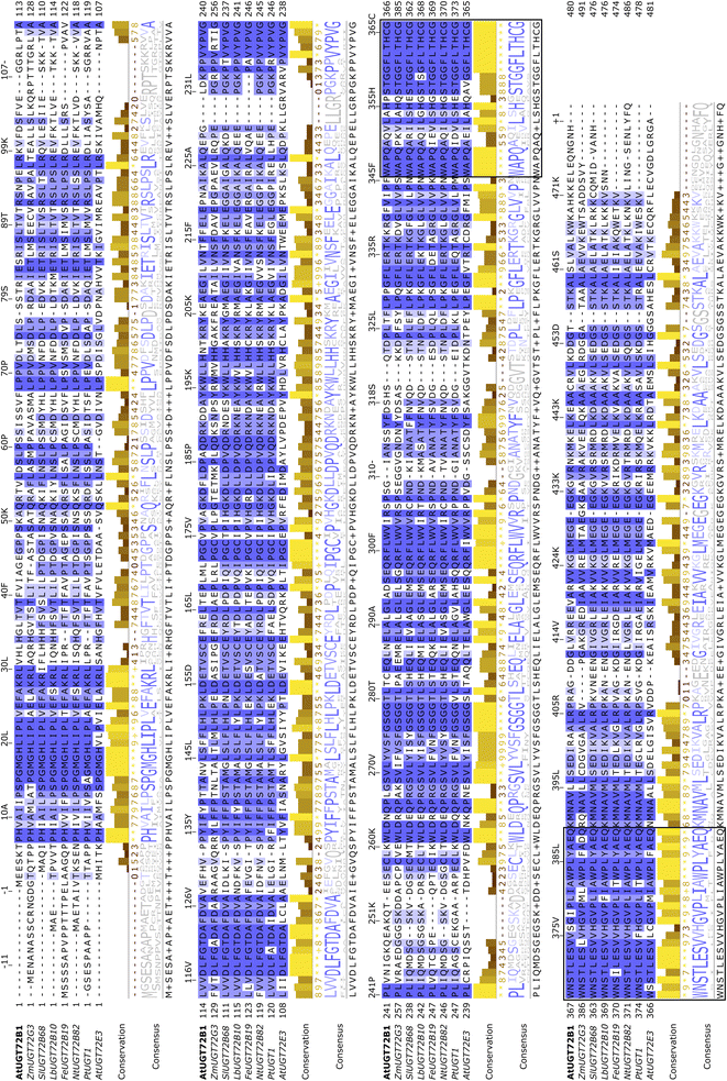 | ||
| Fig. 2 Multiple sequence alignment of the UGTs selected for this study and AtUGT72B1 (in bold). The PSPG motif characteristic of plant UGTs is boxed. | ||
3.2 Glycosylation of 3,4-DCA by the recombinantly expressed UGT72 proteins
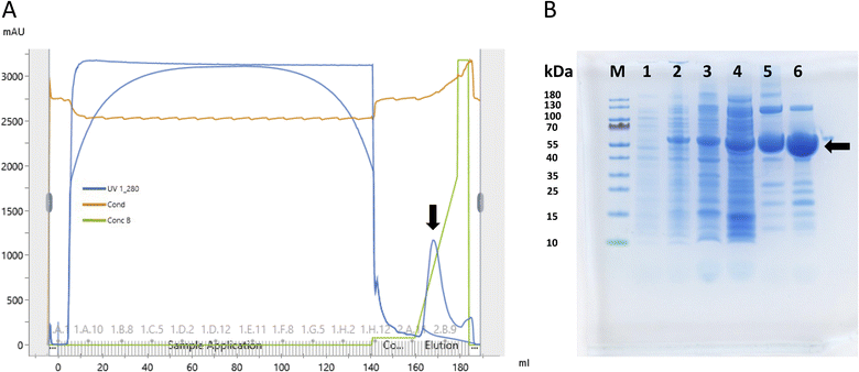
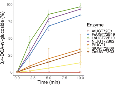
The selected UGTs were expressed in E. coli and purified with nickel-affinity chromatography. The purification results, specifically for LbUGT72B10, are presented in Fig. 3. The partially purified proteins were tested for in vitro activity toward 3,4-DCA. The enzymatic reaction was followed by reverse-phase HPLC and, since an analytical standard for 3,4-DCA-N-glucoside was unavailable, the product of AtUGT72B1 with 3,4-DCA as substrate was used as reference. With the exception of AtUGT72E3, all of the enzymes showed N-glycosylation activity toward 3,4-DCA. Remarkably, LbUGT72B10 demonstrated superior performance, converting almost 50% of 3,4-DCA into 3,4-DCA-N-glucoside within 2 minutes, and achieving nearly 100% conversion yield after 10 minutes (Fig. 4). For this reason, this enzyme was selected for further biochemical and kinetic characterization.3.3 Biochemical and kinetic characterization of LbUGT72B10
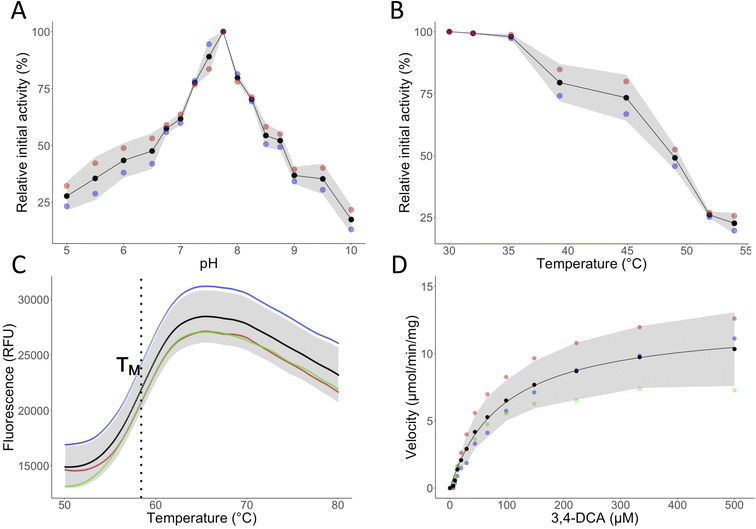
The pH and temperature profiles of LbUGT72B10 were determined with 3,4-DCA as acceptor and UDP-Glc as donor substrates (Fig. 5A and B). LbUGT72B10 was found to have a broad pH tolerance, with activity detected in the pH range of 5.0–9.5 and an optimum pH of 7.75. This result agrees with the published values reported for other UGTs, which are in the range 7.5–9.0.43–48 The temperature optimum was found to be 30 °C, with activity rapidly decreasing beyond 35 °C and no detectable activity at 55 °C. This result conforms to the published values for other UGTs, with their optimal temperature range between 30-40 °C.47–50We further investigated the thermostability of LbUGT72B10 (Fig. 5C). The apparent melting temperature (TM) of UGT72B10 was estimated to be approximately 58.4 ± 0.4 °C using Differential Scanning Fluorimetry (DSF). This is remarkably high for a UGT, which normally melts at approximately 45 °C.37 Although no activity was found beyond 55 °C, this relatively high melting temperature might indicate that LbUGT72B10 possesses superior temporal and chemical stability compared to other UGTs, since these properties often go hand in hand. Our previous study has shown that the reaction conditions to which GT1 enzymes are exposed can impact their thermal stability.37 However, it should be noted that changes in one kind of stability might have an impact on other aspects of stability as well. For example, alterations in pH or temperature can impact conformational and chemical stability, while chemical modifications or denaturation can affect thermal and conformational stability.51–56
After determining the optimal pH and temperature, we proceeded to performing kinetic characterization (Fig. 5D and Table 2). LbUGT72B10 exhibited a comparable catalytic efficiency to that of the previously reported AtUGT72B1.13 However, its kcat is 3-fold higher than that of AtUGT72B1 and 10-fold higher than that of PtUGT1.13,36 On the other hand, the KM of LbUGT72B10 is around three orders of magnitude higher than that of AtUGT72B1.13 Nevertheless, this affinity constant is still considered relatively low when compared to other members of the UGT72 family acting on various acceptors.47,57,58
4. Discussion
Previous studies have suggested that transgenic plants overexpressing specific glycosyltransferases could serve as effective tools for bioremediation.33,38,59 However, our understanding of UGTs involved in the N-glycosylation of 3,4-DCA remains limited, both in vivo and in vitro, and their characterization is lacking. Therefore, this study aimed at expanding our knowledge of UGT-mediated glycosylation of the persistent pollutant 3,4-DCA, since this biological reaction could serve as tool in bioremediation efforts, with the view to expand arable land area and thereby enhance global food security.17 Based on sequence similarity (>60%) to AtUGT72B1, a known glycosylator of 3,4-DCA, we selected seven novel UGT candidates to test for enzymatic activity toward 3,4-DCA. All chosen UGTs had the conserved PSPG motif and the catalytic histidine necessary for N-glycosylation specificity.13,36 All enzymes except AtUGT72E3 showed N-glycosylation activity within 10 minutes, with LbUGT72B10 reaching approximately 100% yield after 10 minutes. LbUGT72B10 was further biochemically and kinetically characterized. The enzyme was active in pH from 5.0 to 9.5, with an optimum pH at 7.75 and a temperature optimum of 30 °C. LbUGT72B10 had a remarkably high melting temperature (58.4 °C), suggesting that the enzyme also possesses chemical stability, as these properties are often interconnected.37,54 These characteristics, together with excellent kinetic properties (full conversion in 10 minutes, KM = 51.2 μM and kcat = 11.2 s−1) align well with the requirements for enzymes employed in various bioremediation strategies.60,61The potential bioremediation approach using transgenic expression of LbUGT72B10 depends on the reproducibility of these in vitro results under in planta conditions, taking into account the influence of the cellular environment on enzyme activity and stability. Also, it is worth noting that currently no genetically modified crops with UGT genes are in use, despite the existence of patented plant UGT sequences for crop protection and bioremediation applications.30 However, even if the in vitro results do not precisely reflect the in vivo context, transgenic plants expressing LbUGT72B10 may still exhibit enhanced resilience toward 3,4-DCA, potentially showing enhanced cellular compartmentalization due to glycosylation, and/or via the higher water solubility of the glycoside promoting efficient 3,4-DCA excretion into the soil, where it can be mineralized by soil microbes.32 3,4-DCA is primarily metabolized through catabolic pathways into naturally occurring intermediates, such as catechol.62 However, only specific microorganisms have been shown to possess 3,4-DCA resistance and to be able to metabolize high concentrations of this xenobiotic, so glycosylation could also mitigate its toxicity toward naturally sensitive microbial populations.63Pichia pastoris, a yeast commonly used in biochemical research which is naturally sensitive to 3,4-DCA, has been shown to survive in the presence of this compound when a UGT, VvUGT72B1, was heterologously expressed in the yeast cells.64 A similar phenomenon may occur in soil microbial populations, if the glucoside rather than the aglycon is excreted in the rhizosphere. Nevertheless, further investigations are required to elucidate the actual bioremediation mechanism in transgenic plants overexpressing LbUGT72B10 and to determine the fate of 3,4-DCA-N-glucoside and its metabolism by soil microorganisms.
5. Conclusions
In conclusion, we have studied seven novel UDP-glycosyltransferases for the glycosylation of the recalcitrant pollutant 3,4-DCA. Among them, LbUGT72B10 from Lycium barbarum exhibited the highest N-glycosylation activity, as well as superior stability and excellent kinetic properties. Although further investigations are needed, the identification of LbUGT72B10 as an efficient enzyme for the glycosylation of 3,4-DCA with potentially superior stability makes it interesting for potential future use in bioremediation efforts of contaminated environments, with the view to expand arable land area and enhance food security and agricultural sustainability.Conflicts of interest
There are no conflicts to declare.Acknowledgements
We thank The Novo Nordisk Foundation for supporting this work through grant NNF20CC0035580.References
- I. Gren, Microbial transformation of xenobiotics, Chemik, 2012, 66(8), 835–842 CAS
.
- C. Leuenberger, W. Giger, R. Coney, J. W. Graydon and E. Molnar-kubica, Persistent chemicals in pulp mill effluents occurrence and behaviour in an activated sludge treatment in plant, Water Res., 1985, 19(7), 885–894 CrossRef CAS
.
- U. G. Ahlborg, T. M. Thunberg and H. C. Spencer, Chlorinated phenols: occurrence, toxicity, metabolism, and environmental impact, Crit. Rev. Toxicol., 1980, 7(1), 1–35 CrossRef CAS PubMed
.
- K. C. Jones and P. De Voogt, Persistent organic pollutants (POPs): state of the science, Environ. Pollut., 1999, 100, 209–221 CrossRef CAS PubMed
.
- S. D. Copley, Diverse mechanistic approaches to difficult chemical transformations: microbial dehalogenation of chlorinated aromatic compounds, Chem. Biol., 1997, 4(3), 169–174 CrossRef CAS PubMed
.
-
M. M. Häggblom and I. D. Bossert, Dehalogenation: Microbial Processes and Environmental Applications, Kluwer Academic Publisher, Boston, 2003 Search PubMed
.
- E. González-Pradas, M. Fernández-Pérez, F. Flores-Céspedes, M. Villafranca-Sánchez, M. D. Ureña-Amate and M. Socías-Viciana,
et al., Effects of dissolved organic carbon on sorption of 3,4-dichloroaniline and 4-bromoaniline in a calcareous soil, Chemosphere, 2005, 59(5), 721–728 CrossRef PubMed
.
- E. Jackson, R. Shoemaker, N. Larian and L. Cassis, Adipose tissue as a site of toxin accumulation, Compr. Physiol., 2017, 7(4), 1085–1135 Search PubMed
.
- M. S. El-Shahawi, A. Hamza, A. S. Bashammakh and W. T. Al-Saggaf, An overview on the accumulation, distribution, transformations, toxicity and analytical methods for the monitoring of persistent organic pollutants, Talanta, 2010, 80(5), 1587–1597 CrossRef CAS PubMed
.
- H. Kraehmer, B. Laber, C. Rosinger and A. Schulz, Herbicides as Weed Control Agents: State of the Art: I. Weed Control Research and Safener Technology: The Path to Modern Agriculture, Plant Physiol., 2014, 166(3), 1119–1131 CrossRef PubMed
.
- W. Aktar, D. Sengupta and A. Chowdhury, Impact of pesticides use in agriculture: their benefits and hazards, Interdiscip. Toxicol., 2009, 2(1), 1–12 Search PubMed
.
- R. Jayaraj, P. Megha and P. Sreedev, Organochlorine pesticides, their toxic effects on living organisms and their fate in the environment, Interdiscip. Toxicol., 2016, 9(3–4), 90–100 CrossRef CAS PubMed
.
- M. Brazier-Hicks, W. A. Offen, M. C. Gershater, T. J. Revett, E. K. Lim and D. J. Bowles,
et al., Characterization and engineering of the bifunctional N-and O-glucosyltransferase involved in xenobiotic metabolism in plants, Proc. Natl. Acad. Sci. U.S.A., 2007, 104(51), 202338–220243 CrossRef PubMed
.
- G. Carvalho, R. Marques, A. R. Lopes, C. Faria, J. P. Noronha and A. Oehmen,
et al., Biological treatment of propanil and 3,4-dichloroaniline: kinetic and microbiological characterisation, Water Res., 2010, 44(17), 4980–4991 CrossRef CAS PubMed
.
- M. Brazier-Hicks, L. A. Edwards and R. Edwards, Selection of plants for roles in phytoremediation: the importance of glucosylation, Plant Biotechnol. J., 2007, 5(5), 627–635 CrossRef CAS PubMed
.
- F. Eissa, F. I. Eissa, A. I. El Makawy, M. I. Badr and O. H. Elhamalawy, Assessment of 3, 4-Dichloroaniline Toxicity as Environmental Pollutant in Male Mice, Eur. J. Biol. Sci., 2012, 4(3), 73–82 Search PubMed
.
- European Chemicals Bureau, European Union Risk Assessment Report 3,4-dichloroaniline (3,4-DCA), 2006, available from http://www.jrc.cec.eu.int Search PubMed.
- A. Claver, P. Ormad, L. Rodríguez and J. L. Ovelleiro, Study of the presence of pesticides in surface waters in the Ebro river basin (Spain), Chemosphere, 2006, 64(9), 1437–1443 CrossRef CAS PubMed
.
- L. M. Games and R. A. Hites, Composition, Treatment Efficiency, and Environmental Significance of Dye Manufacturing Plant Effluents, Anal. Chem., 1997, 49(9), 1433–1439 CrossRef
. Available from:https://pubs.acs.org/sharingguidelines.
- M. Bauchinger, U. Kulka and E. Schmid, Cytogenetic effects of 3,4-dichloroaniline in human lymphocytes and V79 Chinese hamster cells, Mutat. Res., 1989, 226, 197–202 CAS
.
- E. G. Primel, R. Zanella, M. H. S. Kurz, F. F. Gonçalves, M. L. Martins and S. L. O. Machado,
et al., Risk Assessment of Surface Water Contamination by Herbicide Residues: Monitoring of Propanil Degradation in Irrigated Rice Field Waters using HPLC-UV and Confirmation by GC-MS, J. Braz. Chem. Soc., 2007, 18(3), 585–589 CrossRef CAS
.
- A. Elmakawy, A. S. Al-Sarar, Y. Abobakr, F. Eissa, F. I. Eissa and A. I. El Makawy,
et al., Assessment of 3-4 Dichloroaniline Reproductive toxicity and histopathological changes induced by lambda-cyhalothrin in male mice, Eur. J. Biol. Sci., 2012, 4(3), 73–82 Search PubMed
.
- M. A. Ibrahim, S. Z. Zulkifli, M. N. A. Azmai, F. Mohamat-yusuff and A. Ismail, Reproductive toxicity of 3,4-dichloroaniline (3,4-dca) on javanese medaka (oryzias javanicus, bleeker 1854), Animals, 2021, 11(3), 1–12 CrossRef PubMed
.
- M. Bockers, C. Rivero, B. Thiede, T. Jankowski and B. Schmidt, Uptake, Translocation, and Metabolism of 3,4-Dichloroaniline in Soybean and Wheat Plants, Z. Naturforsch., 1994, 49(11–12), 719–726 CrossRef CAS
.
-
D. J. E. R. Cole, Metabolism of Agrochemicals in Plants, ed. Roberts T. R., Wiley, Chichester, UK, 2000, pp. 107–154 Search PubMed
.
- P. M. Coutinho, E. Deleury, G. J. Davies and B. Henrissat, An evolving hierarchical family classification for glycosyltransferases, J. Mol. Biol., 2003, 328(2), 307–317 CrossRef CAS PubMed
.
- D. Bowles, E. K. Lim, B. Poppenberger and F. E. Vaistij, Glycosyltransferases of lipophilic small molecules, Annu. Rev. Plant Biol., 2006, 57, 567–597 CrossRef CAS PubMed
.
- L. L. Lairson, B. Henrissat, G. J. Davies and S. G. Withers, Glycosyltransferases: structures, functions, and mechanisms, Annu. Rev. Biochem., 2008, 77, 521–555 CrossRef CAS PubMed
.
- W. Zhang, S. Wang, J. Yang, C. Kang, L. Huang and L. Guo, Glycosylation of plant secondary metabolites: regulating from chaos to harmony, Environ. Exp. Bot., 2022, 194, 104703 CrossRef CAS
.
- H. Gharabli, V. Della Gala and D. H. Welner, The function of UDP-glycosyltransferases in plants and their possible use in crop protection, Biotechnol. Adv., 2023, 67, 108182 CrossRef CAS PubMed
.
-
M. Brazier-Hicks, W. A. Offen, M. C. Gershater, T. J. Revett, E. K. Lim, D. J. Bowles, et al., Characterization and Engineering of the Bifunctional N-And O-Glucosyltransferase Involved in Xenobiotic Metabolism in Plants, vol. 104, 2007 Search PubMed
.
- L. L. Van Eerd, R. E. Hoagland, R. M. Zablotowicz and J. C. Hall, Pesticide metabolism in plants and microorganisms, Weed Sci., 2003, 51, 472–495 CrossRef CAS
.
- C. Loutre, D. P. Dixon, M. Brazier, M. Slater, D. J. Cole and R. Edwards, Isolation of a glucosyltransferase from Arabidopsis thaliana active in the metabolism of the persistent pollutant 3,4-dichloroaniline, Plant J., 2003, 34, 485–493 CrossRef CAS PubMed
.
- T. Louveau, A. Orme, H. Pfalzgraf, M. J. Stephenson, R. Melton and G. Saalbach,
et al., Analysis of two new arabinosyltransferases belonging to the carbohydrate-active enzyme (CAZY) glycosyl transferase family1 provides insights into disease resistance and sugar donor specificity, Plant Cell, 2018, 30(12), 3038–3057 CrossRef CAS PubMed
.
- J. Huang, J. Li, J. Yue, Z. Huang, L. Zhang and W. Yao,
et al., Functional Characterization of a Novel Glycosyltransferase (UGT73CD1) from Iris tectorum Maxim. for the Substrate promiscuity, Mol. Biotechnol., 2021, 63(11), 1030–1039 CrossRef CAS PubMed
.
- D. Teze, J. Coines, F. Fredslund, K. D. Dubey, G. N. Bidart and P. D. Adams,
et al., O-/N-/S-Specificity in Glycosyltransferase Catalysis: From Mechanistic Understanding to Engineering, ACS Catal., 2021, 11(3), 1810–1815 CrossRef CAS
.
- D. Teze, G. N. Bidart and D. H. Welner, Family 1 glycosyltransferases (GT1, UGTs) are subject to dilution-induced inactivation and low chemo stability toward their own acceptor substrates, Front. Mol. Biosci., 2022, 9 Search PubMed
.
- B. Meßner, O. Thulke and A. R. Schäffner, Arabidopsis glucosyltransferases with activities toward both endogenous and xenobiotic substrates, Planta, 2003, 217(1), 138–146 CrossRef PubMed
.
- S. F. Altschup, W. Gish, W. Miller, E. W. Myers and D. J. Lipman, Basic Local Alignment Search Tool, J. Mol. Biol., 1990, 215, 403–410 CrossRef PubMed
.
- N. Speeckaert, M. El Jaziri, M. Baucher and M. Behr, UGT72, a Major Glycosyltransferase Family for Flavonoid and Monolignol Homeostasis in Plants, Biology, 2022, 11(3), 441 CrossRef CAS PubMed
.
- S. A. Osmani, S. Bak, A. Imberty, C. E. Olsen and B. L. Møller, Catalytic key amino acids and UDP-sugar donor specificity of a plant glucuronosyltransferase, UGT94B1: Molecular modeling substantiated by site-specific mutagenesis and biochemical analyses, Plant Physiol., 2008, 148(3), 1295–1308 CrossRef CAS PubMed
.
- J. Hughes and M. A. Hughes, Multiple secondary plant product UDP-glucose glucosyltransferase genes expressed in cassava (manihot esculenta crantz) cotyledons, DNA Sequence, 1994, 5(1), 41–49 CrossRef CAS PubMed
.
- C. M. Ford, P. K. Boss and P. Bordier Høj, Cloning and Characterization of Vitis vinifera UDP-Glucose:Flavonoid 3-O-Glucosyltransferase, a Homologue of the Enzyme Encoded by the Maize Bronze-1 Locus That May Primarily Serve to Glucosylate Anthocyanidins in Vivo, J. Biol. Chem., 1998, 273(15), 9224–9233 CrossRef CAS PubMed
.
- C. Paczkowski, M. Kalinowska and Z. A. Wojciechowski, UDP-glucose:solasodine glucosyltransferase from eggplant (Solanum melongena L.) leaves: partial purification and characterization, Acta Biochim. Pol., 1997, 44(1), 43–54 CrossRef CAS PubMed
.
- A. D. Parry and R. Edwards, Characterization of O-glucosyltransferases with activities toward phenolic substrates in alfalfa, Phytochemistry, 1994, 37(3), 655–661 CrossRef CAS
.
- R. L. Durren and C. A. Mcintosh, Flavanone-7-O-glucosyltransferase activity from Petunia hybrida, Phytochemistry, 1999, 52, 793–798 CrossRef CAS PubMed
.
- Y. Tan, J. Yang, Y. Jiang, J. Wang, Y. Liu and Y. Zhao,
et al., Functional Characterization of UDP-Glycosyltransferases Involved in Anti-viral Lignan Glycosides Biosynthesis in Isatis indigotica, Front. Plant Sci., 2022, 13 Search PubMed
.
- K. Ghose, K. Selvaraj, J. McCallum, C. W. Kirby, M. Sweeney-Nixon and S. J. Cloutier,
et al., Identification and functional characterization of a flax UDP-glycosyltransferase glucosylating secoisolariciresinol (SECO) into secoisolariciresinol monoglucoside (SMG) and diglucoside (SDG), BMC Plant Biol., 2014,(1), 14 Search PubMed
.
- P. Srivastava, A. Garg, R. C. Misra, C. S. Chanotiya and S. Ghosh, UGT86C11 is a novel plant UDP-glycosyltransferase involved in labdane diterpene biosynthesis, J. Biol. Chem., 2021, 297(3), 101045 CrossRef CAS PubMed
.
- B. Wu, X. Cao, H. Liu, C. Zhu, H. Klee and B. Zhang,
et al., UDP-glucosyltransferase PpUGT85A2 controls volatile glycosylation in peach, J. Exp. Bot., 2019, 70(3), 985–994 CrossRef PubMed
.
- R. M. Daniel, The upper limits of enzyme thermal stability, Enzyme Microb. Technol., 1996, 19, 74–79 CrossRef CAS
.
- B. M. Beadle and B. K. Shoichet, Structural bases of stability-function tradeoffs in enzymes, J. Mol. Biol., 2002, 321(2), 285–296 CrossRef CAS PubMed
.
- A. S. Panja, S. Maiti and B. Bandyopadhyay, Protein stability governed by its structural plasticity is inferred by physicochemical factors and salt bridges, Sci. Rep., 2020, 10(1), 1822 CrossRef CAS PubMed
.
- A. Razvi and J. M. Scholtz, Lessons in stability from thermophilic proteins, Protein Sci., 2006, 15(7), 1569–1578 CrossRef CAS PubMed
.
- K. Singh, M. Shandilya, S. Kundu and A. M. Kayastha, Heat, acid and chemically induced unfolding pathways, conformational stability and structure-function relationship in wheat α-amylase, PLoS One, 2015, 10(6), 1–18 Search PubMed
.
- G. Bidart, D. Teze, C. Jansen, E. Pasutto, N. Putkaradze, A. M. Sesay, et al., Chemoenzymatic indican for sustainable light-driven denim dyeing, 2023, PREPRINT (Version 1) available at Research Square, https://doi.org/10.21203/rs.3.rs-2416810/v1 Search PubMed.
- Q. Yin, G. Shen, Z. Chang, Y. Tang, H. Gao and Y. Pang, Involvement of three putative glucosyltransferases from the UGT72 family in flavonol glucoside/rhamnoside biosynthesis in Lotus japonicus seeds, J. Exp. Bot., 2017, 68(3), 597–612 CAS
.
- G. Sun, J. Liao, E. Kurze, T. D. Hoffmann, W. Steinchen and K. McGraphery,
et al., Apocarotenoids are allosteric effectors of a dimeric plant glycosyltransferase involved in defense and lignin formation, New Phytol., 2023, 238, 2080–2098 CrossRef CAS PubMed
.
- E. K. Lim and D. J. Bowles, A class of plant glycosyltransferases involved in cellular homeostatis, EMBO J., 2004, 23, 2915–2922 CrossRef CAS PubMed
.
- T. D. Sutherland, I. Horne, K. M. Weir, C. W. Coppin, M. R. Williams and M. Selleck,
et al., Enzymatic bioremediation: from enzyme discovery to applications, Clin. Exp. Pharmacol. Physiol., 2004, 31(11), 817–821 CrossRef CAS PubMed
.
- C. Scott, G. Pandey, C. J. Hartley, C. J. Jackson, M. J. Cheesman and M. C. Taylor,
et al., The enzymatic basis for pesticide bioremediation, Indian J. Microbiol., 2008, 48, 65–79 CrossRef CAS PubMed
.
- A. Wasserfallen, J. Zeyer and K. N. Timmis, Bacterial metabolism and toxicity of halogenated anilines, Experientia, 1986, 42, 106 CrossRef
.
- A. L. Tasca and A. Fletcher, State of the art of the environmental behaviour and removal techniques of the endocrine disruptor 3,4-dichloroaniline, J. Environ. Sci. Health, Part A, 2018, 53(3), 260–270 CrossRef CAS PubMed
.
- Z. S. Xu, W. Xue, A. S. Xiong, Y. Q. Lin, J. Xu and B. Zhu,
et al., Characterization of a bifunctional O- and N-glucosyltransferase from Vitis vinifera in glucosylating phenolic compounds and 3,4-dichloroaniline in Pichia pastoris and Arabidopsis thaliana, PLoS One, 2013, 8(11), e80449 CrossRef PubMed
.
| This journal is © The Royal Society of Chemistry 2023 |

