
 Open Access Article
 This Open Access Article is licensed under a Creative Commons Attribution-Non Commercial 3.0 Unported Licence
Efficient photoactivated hydrogen evolution promoted by CuxO–gCN–TiO2–Au (x = 1,2) nanoarchitectures†
Mattia Benedet ab,
Gian Andrea Rizziab,
Alberto Gasparottoab,
Lunjie Zengc,
Gioele Pagotd,
Eva Olssonc,
Vito Di Notod,
Chiara Maccato*ab and
Davide Barrecab
ab,
Gian Andrea Rizziab,
Alberto Gasparottoab,
Lunjie Zengc,
Gioele Pagotd,
Eva Olssonc,
Vito Di Notod,
Chiara Maccato*ab and
Davide Barrecab
aDepartment of Chemical Sciences, Padova University, INSTM, 35131 Padova, Italy. E-mail: chiara.maccato@unipd.it
bCNR-ICMATE, INSTM, Department of Chemical Sciences, Padova University, 35131 Padova, Italy
cDepartment of Physics, Chalmers University of Technology, 41296 Gothenburg, Sweden
dSection of Chemistry for the Technology (ChemTech), Department of Industrial Engineering, Padova University, INSTM, 35131 Padova, Italy
First published on 28th February 2024
Abstract
In this work, we propose an original and potentially scalable synthetic route for the fabrication of CuxO–gCN–TiO2–Au (x = 1,2) nanoarchitectures, based on Cu foam anodization, graphitic carbon nitride liquid-phase deposition, and TiO2/Au sputtering. A thorough chemico-physical characterization by complementary analytical tools revealed the formation of nanoarchitectures featuring an intimate contact between the system components and a high dispersion of gold nanoparticles. Modulation of single component interplay yielded excellent functional performances in photoactivated hydrogen evolution, corresponding to a photocurrent of ≈−5.7 mA cm−2 at 0.0 V vs. the reversible hydrogen electrode (RHE). These features, along with the very good service life, represent a cornerstone for the conversion of natural resources, as water and largely available sunlight, into added-value solar fuels.
1 Introduction
The progressively higher global demand of energy, whose supply is still predominantly dependent on fossil fuels, and the resulting climate changes aggravation are the motivating forces for numerous efforts aimed at achieving alternative and sustainable solutions.1–7 In this regard, sunlight-driven water splitting to yield molecular hydrogen (H2), a zero-emission and renewable energy vector, stands as a strategic choice for the conversion of an inexhaustible natural resource into chemical energy,8–17 provided that a careful selection and engineering of active semiconductor photoelectrocatalysts is performed.7,10,12,18,19 Among the possible candidates for the hydrogen evolution reaction (HER), an effective and spotless process to deliver hydrogen,5 p-type copper oxides (CuxO, x = 1,2; Eg ≈ 2.1 and ≈1.5 eV for x = 1 and 2, respectively) are inexpensive, non-toxic and naturally abundant semiconductors featuring efficient sunlight harvesting.1,10,12,20–23 In particular, CuO has also been proposed as a Cu2O overlayer to achieve an improved charge carrier transport towards the electrolyte, suppressing the detrimental recombination of photogenerated electrons and holes. In addition, CuO/Cu2O systems feature an extended spectral absorption compared to Cu2O,2,13,14 allowing thus an enhanced harvesting of the incoming electromagnetic radiation.To further improve charge transfer kinetics and overcome instability problems due to photocorrosion,1,14,16,17,23 different literature works have so far been focused on the ad hoc modification of CuxO surface with adequate partners, to yield nanoheterojunctions providing supplementary benefits.1,13,20 To this aim, graphitic carbon nitride (gCN), a burgeoning n-type semiconductor possessing a two-dimensional structure (Eg ≈ 2.7 eV), presents several concurrent advantages, including cost effectiveness, eco-friendly character, stability, tuneable electronic structure, and attractive photoactivity.4,11,19,24–27 The system performances can be further improved by the additional introduction of oxide semiconductors and/or metals, obtaining multi-component functional nanoarchitectures.9,12 In particular, TiO2 can exert a protective action against CuxO photocorrosion, limit the undesired recombination of photocarriers affecting gCN, and boost charge mobility at the photoelectrocatalyst–electrolyte interface.3,8,15,17,18,21,28,29 The latter advantage can be favourably amplified by functionalizing the target systems with low amounts of optimally dispersed Au nanoparticles (NPs), acting as electric/catalytic/optical mediators for the achievement of efficient H2 generation.9,15,23,25,26
In the present study, we propose for the first time the fabrication of CuxO–gCN–TiO2–Au (x = 1,2) nanoarchitectures through an original multi-step synthetic strategy (Fig. 1a; see also the ESI†). Metallic Cu foams, offering high porosity and electrical conductivity, as well as electrolyte accessibility and efficient release of gaseous products,6,30 are initially subjected to anodization and subsequent thermal treatment in air for the in situ growth of CuxO.22,31 The proposed route affords quasi-1D nanostructures with a high active area, promoting, in turn, a homogeneous distribution of gCN, whose deposition was carried out by dipping in urea-containing solutions, followed by annealing.10 The eventual introduction of TiO2 and Au NPs was performed by sequential radio frequency (RF)-sputtering, carried out under mild conditions to avoid alterations of the underlying system nano-organization. The target specimens are characterized by an enhanced mechanical stability in comparison to electrocatalysts obtained by immobilization of inks/pastes with additives/binders on various substrates.4,5,7,11,19,28 In addition, the synergy between the RF-sputtering infiltration power and the high area morphology30,32 of the pristine CuxO–gCN enabled an intimate contact between the single components, and an efficient dispersion of Au NPs. Altogether, these features yielded, for CuxO–gCN–TiO2–Au photoelectrocatalysts, a photocurrent density of ≈−5.7 mA cm−2 at 0.0 V vs. RHE and a Tafel slope of ≈−160 mV dec−1, among the best ever reported in the literature up to date for homologous CuxO- and/or gCN-containing systems in such applications.
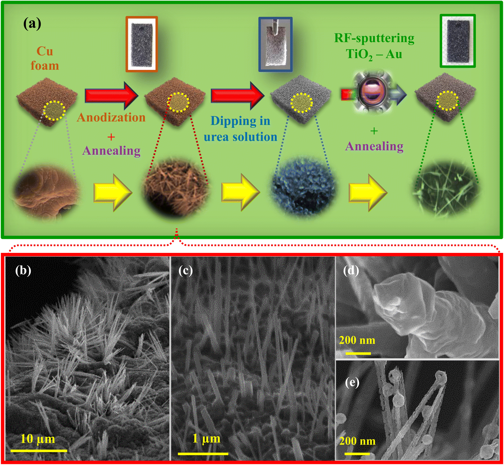 | ||
| Fig. 1 (a) Sketch of the preparation route to CuxO–gCN–TiO2–Au (x = 1,2) photoelectrocatalysts used in the present work. (b–e) Representative field emission-scanning electron microscopy (FE-SEM) micrographs for CuxO systems. | ||
2 Experimental
2.1 Material preparation
2.2 Characterization
FE-SEM images were collected with a Zeiss Supra 40 VP instrument equipped with an INCA x-act PentaFET Precision spectrometer for energy-dispersive X-ray spectroscopy (EDXS), using primary beam voltages between 10 and 20 kV. X-ray photoelectron spectroscopy (XPS) analyses were carried out with a Specs EnviroESCA apparatus equipped with an AlKα X-ray source (hν = 1486.6 eV). Binding energy (BE) values (uncertainty = ±0.1 eV) were corrected for charging phenomena by assigning a position of 284.8 eV to the C 1s signal arising from adventitious contamination.34 Peak fitting was performed using the Keystone software (Specs) and applying a Shirley-type background subtraction.A JEOL monochromated ARM200F instrument, operated at 200 kV, was used for scanning transmission electron microscopy (STEM) imaging, STEM-EDXS and STEM-electron energy loss spectroscopy (EELS) analyses. The apparatus was equipped with a double-Wien type monochromator, CEOS probe and image Cs correctors, a double silicon drift detector (SDD) for EDXS, and a Gatan image filter (GIF) Continuum for EELS. STEM annular dark field (ADF) images were recorded with beam convergence and inner collection half-angles of ≈27 mrad and ≈50 mrad, respectively.
The system photoelectrocatalytic activity was tested in 0.5 M KHCO3 aqueous solution (Carlo Erba, pH = 8.5), using an Autolab PGSTAT204 potentiostat/galvanostat. The fabricated materials were used as working electrodes, whereas a standard Hg/HgO (MMO) electrode and a Pt wire were used as reference and counter-electrodes, respectively. During each test, the sample was illuminated from the front side (electrode–electrolyte interface) using a white light LED source (intensity ≈ 100 mW cm−2).
Additional details on material preparation and characterization are available in the ESI.†
3 Results and discussion
The system morphology and its interplay with the processing conditions were initially investigated by FE-SEM. As revealed by Fig. 1b–e, the Cu foam anodization and subsequent annealing yielded high area arrays of copper oxide nanowires (average aspect ratio ≈5000 nm/150 nm = 33), some of which featured the presence of spheres (diameter ≈ 100 nm) on their tips. The obtainment of such structures is not controlled by vapor–solid or vapor–liquid–solid mechanisms,35 but depends on Cu oxidation at the electrode/electrolyte interface during the anodization step, and subsequent CuxO nanowires formation upon annealing, as reported by previous investigators.36–39 FE-SEM images and EDXS results for composite systems, along with pertaining observations, are reported in the ESI (see Fig. S2, S3 and Page S6).†X-ray diffraction (XRD) measurements for all the target specimens revealed the co-presence of Cu2O and CuO, along with reflections of metallic copper arising from the Cu foam substrate (Fig. S4†). No appreciable signals originating from gCN, TiO2, or Au could ever be detected. The lack of diffraction peaks attributable to graphitic carbon nitride was due to its amorphous character (see also below), whereas the absence of titanium dioxide and gold reflections could be traced back to their low amount and high dispersion. On the other hand, gCN, Au and TiO2 presence was demonstrated by XPS, TEM and related outcomes (see below).
Additional key information on the system chemical composition and elemental oxidation states was provided by XPS analyses (see also Fig. S5†). As a matter of fact, titanium and gold photopeaks were undetectable (see Fig. S6a and b†), indicating thus that TiO2 and Au were not present in the outermost material regions (compare TEM data and pertaining observations). Irrespective of the analyzed sample, the Cu 2p peak shape and position [Fig. 2a; BE (Cu 2p3/2) ≈933.8 eV], along with the presence of shake-up satellites, agreed with CuO presence.1,6,8,10,21,23 Evaluation of copper Auger parameters (see also the ESI and Fig. S6c†) yielded α = 1851.4 eV, in agreement with the surface presence of CuO.34,40–43 The O/Cu atomic percentage (at%) ratio values (1.5–1.6) were higher than the stoichiometric one (1.0) expected for CuO, suggesting the presence of chemisorbed –OH groups and adsorbed H2O on the system surface. Accordingly, O 1s signals (Fig. 2b) resulted from the concurrence of CuO lattice oxygen (I, BE = 529.9 eV),21,23 surface chemisorbed –OH groups (II, BE = 531.5 eV),3,25 and adsorbed water/C–O bonds between gCN and CuO (both contributing to band III, located at BE = 533.0 eV).10,24 Indeed, –OH presence is beneficial for the target end-use,1,21 and gCN–CuO bonding is favourable to obtain an improved electron–hole separation,24 resulting thus in higher functional performances. The C 1s signal (Fig. 2c), beside the adventitious carbon band (IV, BE = 284.8 eV),3,9,16,25,28 presented a component at ≈286.1 eV (V) due to both C–O bonds and −C−NHx moieties (x = 1,2) on gCN heptazine ring edges.3,4,17,24,44 Finally, signal VI at BE ≈ 288.1 eV was attributed to N–C![[double bond, length as m-dash]](https://www.rsc.org/images/entities/char_e001.gif) N carbon atoms in gCN.1,8,16,18,28 In line with these results, N 1s components (Fig. 2d) were related to bi-coordinated nitrogen (CN–C; VII, BE = 398.7 eV),4,8,24,28 tertiary N–(C)3 atoms (VIII, BE = 399.9 eV),1,8,24,26,28 and –NHx groups (IX, BE = 401.1 eV).3,16,28,45
N carbon atoms in gCN.1,8,16,18,28 In line with these results, N 1s components (Fig. 2d) were related to bi-coordinated nitrogen (CN–C; VII, BE = 398.7 eV),4,8,24,28 tertiary N–(C)3 atoms (VIII, BE = 399.9 eV),1,8,24,26,28 and –NHx groups (IX, BE = 401.1 eV).3,16,28,45
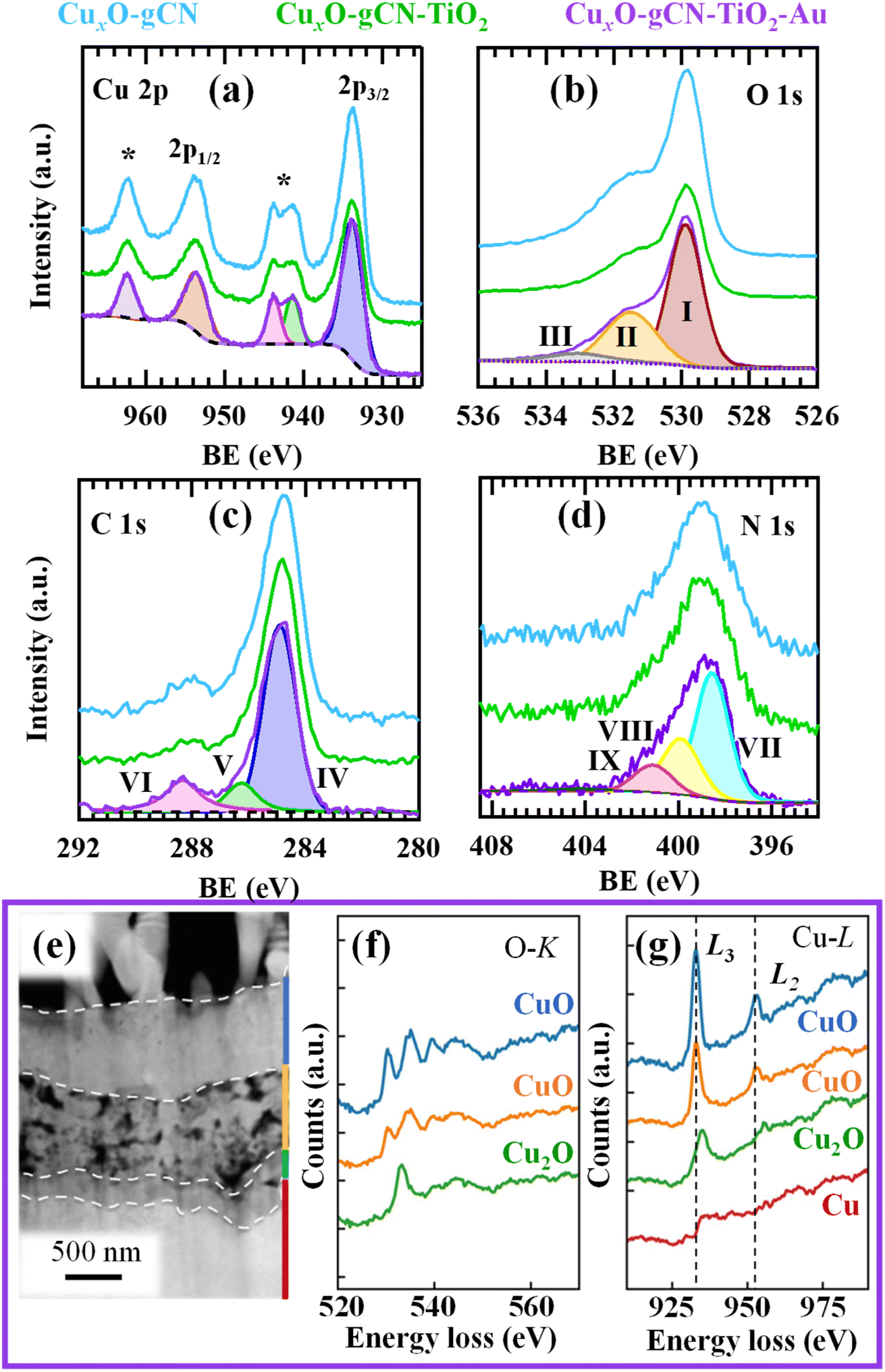 | ||
| Fig. 2 Cu 2p (a), O 1s (b), C 1s (c), and N 1s (d) photoelectron peaks for CuxO–gCN, CuxO–gCN–TiO2, and CuxO–gCN–TiO2–Au. For the latter sample, fit components are displayed along with raw spectra as a representative case. In (a), shake-up satellites are denoted by *. (e) Representative cross-sectional STEM-ADF image for CuxO–gCN–TiO2–Au. Dashed lines mark interfaces between adjacent layers; the middle one is porous, while the others are continuous. The average thickness is ≈1 μm, 1.5 μm, and 200 nm for the top (blue), middle (orange) and bottom (green) layers, respectively. (f and g) Corresponding electron energy loss spectroscopy (EELS) spectra of O-K and Cu-L edges. The positions of Cu L3,2 white-lines are marked by dashed lines. | ||
An important insight into the system nano-organization was gained by TEM and related analyses. Preliminary STEM imaging on CuxO–gCN–TiO2–Au (Fig. 2e) revealed the presence of a stacked structure over the Cu foam, mainly composed of Cu and O, and characterized by diverse layers showing different Cu–O bonding features, as demonstrated by EELS results. In fact, the energy loss near edge structure (ELNES) in EELS is sensitive to variations in local chemical bonding,46 i.e. in copper valence and coordination.44 In the top two layers (marked in blue and orange in Fig. 2e), the features of O-K and Cu-L ELNES signals supported the presence of CuO,44 whereas the O-K ELNES of the layer next to Cu substrate (marked in green in Fig. 2e) indicates the occurrence of Cu2O. The O-K top layer spectrum presented four signals located between ≈530 and 550 eV (Fig. 2f), whereas only an intense peak (530–540 eV) and a broad one (540–550 eV) could be observed for the bottom layer. As regards Cu-L edge, a slight shift in L3 and L2 positions occurred between top and bottom spectra (Fig. 2g), accompanied by a L3/L2 intensity ratio variation. Taken together, these results indicated that the bottom layer consists of Cu2O, while the porous middle layer possesses fine structures similar to the top CuO one. Additional observations highlighted that Au NPs, mostly found in the porous copper oxide region (Fig. 3a and b), which is not on the top of the target specimen, were single-crystalline and free from any obvious clustering. As can be observed in Fig. 3c and d, their structure was consistent with that of metallic gold. EDXS chemical maps (Fig. 3e–j) demonstrated that, whereas Au NPs were dispersed within copper oxide, titanium dioxide was concentrated in relatively small regions, forming strip-shaped domains in close vicinity to Au nanoparticles (Fig. 3i and j; see also Fig. S7†). In agreement with XRD outcomes, carbon nitride turned out to be amorphous in the target specimens (see Fig. S8†).
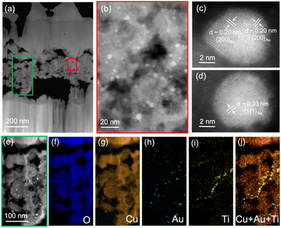 | ||
| Fig. 3 (a) Overview cross-sectional STEM-ADF image for CuxO–gCN–TiO2–Au. (b) STEM-ADF micrograph of the area marked by the red window in (a). Au nanoparticles (dimensions = 2÷10 nm) show a higher contrast than CuxO and a high image intensity, due to the relatively large atomic number of gold. (c and d) Atomic resolution STEM-ADF images of Au NPs, with indexing of gold lattice fringes and pertaining interplanar distances. The lattice fringes below the Au nanoparticle in (d) are likely coming from TiO2. (e) STEM-ADF image of the area marked by the green box in (a), and corresponding STEM-EDXS elemental maps of O (f), Cu (g), Au (h), Ti (i), as well as the superimposed map of copper, gold, and titanium (j). | ||
As a matter of fact, the open morphology of the underlying copper foam substrate, along with the peculiar features of CuxO (consisting of a 3D array of nanowires protruding from the foam meshes) resulted in a very porous CuxO 3D scaffold, promoting a very close contact between the single material constituents.30 In this regard, Au and TiO2 in-depth dispersion was further enhanced by the inherent infiltration power of the sputtering technique,48,49 that also triggers the diffusion of sputtered species into the innermost Cu/CuxO regions thanks to the continuous plasma bombardment of the growing material.50 The synergistic concurrence of these effects, along with the (relatively) small gold and titanium dioxide amounts, resulted in the dispersion of such species in sub-surface regions that cannot be sampled by XPS, and require in fact alternative analytical techniques, as TEM, with local-probing capability on a nanometer-size scale.
The above discussed intimate contact between the system constituents is a key issue enabling to benefit from their synergistic interplay, as showed by functional HER tests (see Fig. 4, S9, and S10†). In fact, a photocurrent density enhancement took place upon CuxO functionalization with gCN, TiO2, and Au [Fig. 4a and b; see also applied bias photon-to-current efficiency (ABPE) curves in Fig. 4c] yielding, for CuxO–gCN–TiO2–Au, a remarkable value of ≈−5.7 mA cm−2 at 0.0 V vs. RHE. Such a very attractive result can be attributed to the successful formation of a high density of heterojunctions, providing various concurrent advantages: (i) a protective action towards Cu2O, suppressing its dismutation to Cu and CuO under illumination;2,13,19,51 (ii) an improved separation of photogenerated charge carriers, resulting in a confinement of holes in Cu2O VB and a transfer of electrons into the CBs of the other photoelectrocatalyst constituents up to the interface with the electrolyte.12,17,22,52,53 As a consequence, attractive performances could be achieved. In fact, the HER photocurrent onset occurred at higher potentials with respect to bare CuxO as soon as the junction with gCN was formed (0.32 V), and underwent further improvements after the introduction of titanium dioxide (0.34 V) and gold (0.36 V; Fig. 4d). In particular, the most active photoelectrocatalyst turned out to be CuxO–gCN–TiO2–Au, for which the band structure arrangement yielded the best electron–hole separation. The resulting energy level alignment (Fig. 4e) match well with the positions of flat band potentials obtained by Mott–Schottky plots (Fig. S11†). Upon irradiation, the system components undergoing excitation are Cu2O, CuO, and gCN, whereas Au NPs work as electron sinks with improvement of the electrical contact. The introduction of titanium dioxide, which is not excited by the adopted light source (see also Fig. S1†), exerted a protective action against photocorrosion, and enabled an improvement of photogenerated electron–hole separation, facilitating charge injection into the electrolyte.
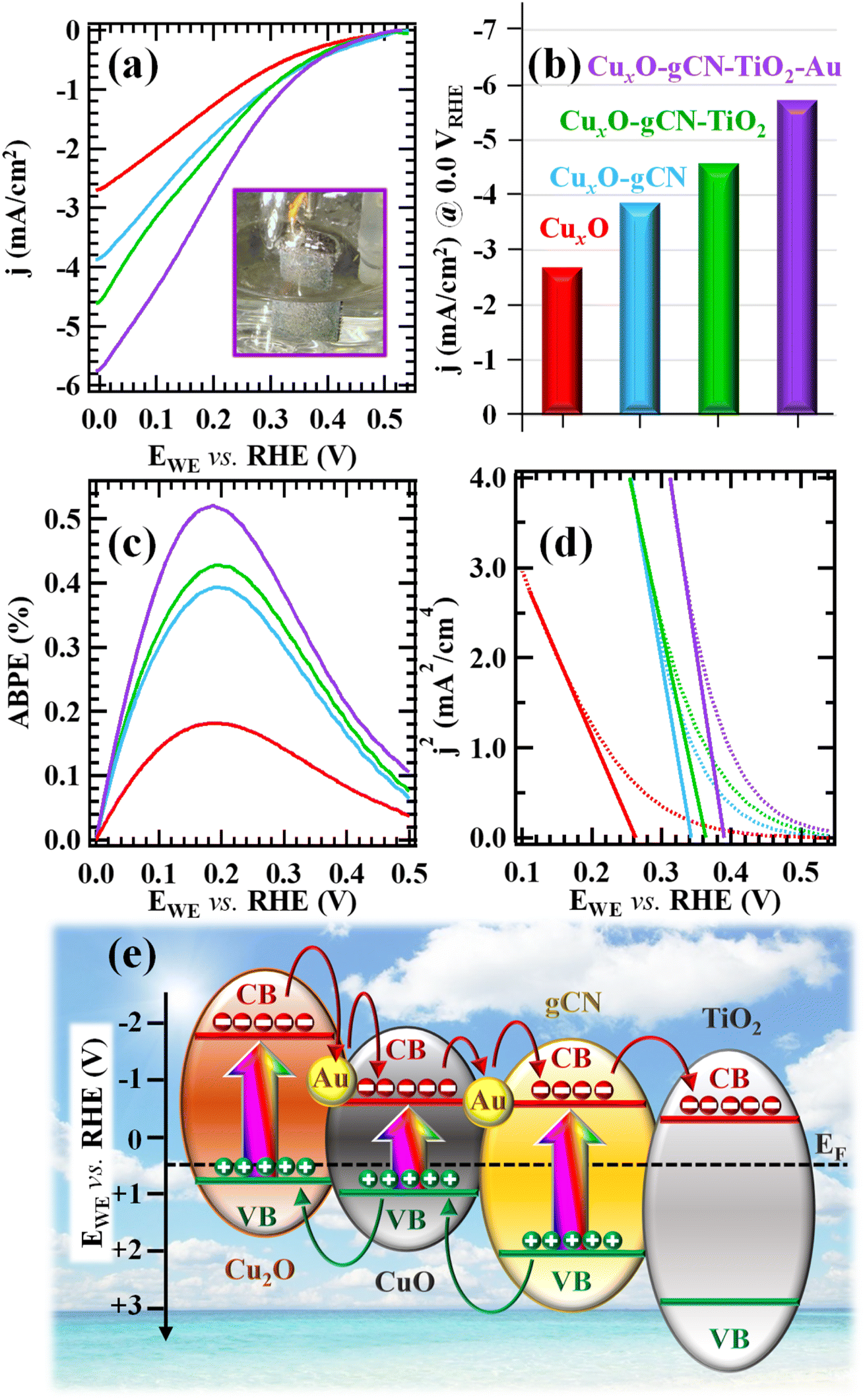 | ||
| Fig. 4 Electrochemical characterization of the target photocathodes. (a) Linear-sweep voltammetry (LSV) plots of photocurrent vs. applied potential. Inset: Image of CuxO–gCN–TiO2–Au during the LSV up to 0.0 V vs. RHE, evidencing the formation of H2 bubbles. (b) Bar diagram reporting photocurrent densities at 0.0 V. (c) ABPE (%) curves. (d) Square photocurrent density (j2) vs. bias curves, for Fermi level (EF) extrapolation through the intercept with the potential axis.47 (e) Schematic representation of the band structure arrangement for CuxO–gCN–TiO2–Au. CB and VB indicate conduction and valence band edges, respectively. | ||
The positive activity improvement observed upon going from CuxO to CuxO–gCN–TiO2–Au was also confirmed by Tafel slopes (yielding, for the best performing CuxO–gCN–TiO2–Au specimen, a value of ≈−160 mV dec−1; see Fig. S12†). Interestingly, the presently reported HER performances compare very favourably with those of numerous homologous electrocatalysts previously reported in the literature up to date (see Tables S1 and S2†). These characteristics, related to the unique material morphological and compositional features (see above and ESI†), were accompanied by a very good operational stability (see Fig. S13 and S14†). This conclusion was also corroborated by post-operando XPS analyses, allowing to exclude the occurrence of major compositional changes upon repeated photoelectrochemical testing. In fact, neither the main photoelectron peaks (Fig. S15†; compare with Fig. 2a–d and S6†), nor quantitative analyses results (Fig. S16†) exhibited significant differences with respect to the original situation, suggesting that, differently from previously reported results,12 no appreciable CuO conversion into Cu2O and metallic Cu took place over time. In accordance with these outcomes, FE-SEM measurements (Fig. S17†; compare with Fig. S2c†) revealed that the system morphology did not appreciably change after functional tests, enabling thus to rule out significant degradation phenomena under operation. In perspective, this behaviour paves the way to further implementations for possible real-world end-uses, as also indicated by a preliminary proof-of-concept test aimed at bias-free photoactivated H2 production for CuxO–gCN–TiO2–Au, the best performing specimen in HER experiments. As can be observed in Fig. S18a,† after an initial induction period of ≈2 h, H2 yield underwent a net increase and subsequently remained almost constant for irradiation times >20 h (mean value ≈1.8 mmol h−1 m−2). This result ruled out the existence of photocorrosion processes, and highlighted an appreciable material service life. The latter feature, of notable importance for eventual practical applications,32,54 was further confirmed by the linear trend of Fig. S18b.† The favorable activity in comparison with homologous photocatalysts, and even of Pt-containing ones (see the ESI, Pages S25–S26†), and the absence of platinum in the present supported nanosystems, that contain a minimal amount of active material,32 render the target catalysts interesting platforms for future technological developments.
4 Conclusions
In conclusion, this work has reported on a modular and scalable route for the preparation of green CuxO–gCN–TiO2–Au (x = 1,2) nanoarchitectures on Cu foam substrates. In particular, after the initial anodization of metallic copper foams and the liquid-phase introduction of graphitic carbon nitride, the system functionalization with titanium dioxide and gold has been accomplished by means of RF-sputtering. A careful selection of the adopted operational conditions yielded high area, multicomponent electrocatalysts, benefitting from the intimate contact of the single constituents. The synergistic chemical and electronic interplay between the latter enabled to obtain a remarkable HER activity in terms of photocurrent densities and Tafel slopes, accompanied by a very good stability. The insights provided by the present study could act as a pointer to master tailored nanoheterostructures for energy-related applications, with particular regard to the engineering of high performance photocathodes for large-scale production of green hydrogen via solar-activated water electrolysis. Based on the present results, optimization of material performances and detailed studies on bias-free photocatalytic hydrogen evolution from aqueous media will also be performed, in order to attain a detailed insight into the interplay between material composition and the resulting photoactivity for practical end-uses.Author contributions
Mattia Benedet: data curation, visualization, and writing – original draft; Gian Andrea Rizzi: formal analysis, investigation and validation; Alberto Gasparotto: data analysis and validation; Lunjie Zeng: data curation and visualization; Gioele Pagot: formal analysis and investigation; Eva Olsson: funding acquisition, and writing – review & editing; Vito Di Noto: formal analysis, and writing – review & editing; Chiara Maccato: conceptualization, validation, supervision, funding acquisition, and writing – review & editing; Davide Barreca: supervision, funding acquisition, and writing.Conflicts of interest
There are no conflicts to declare.Acknowledgements
The present work was financially supported by Progetti di Ricerca@CNR – avviso 2020 – ASSIST, Padova University (P-DiSC#04BIRD2020-UNIPD EUREKA, P-DiSC#02BIRD2023-UNIPD RIGENERA, DOR 2021–2023), INSTM Consortium (INSTM21PDGASPAROTTO-NANOMAT, INSTM21PDBARMAC-ATENA), Supersonic Cluster beam synthesis of Innovative TRansition metal Oxides PHotoelectrodes for HYdrogen production (SCI-TROPHY) PRIN 2022 2022474YE8, CUP J53D23007340008, and the European Union’s Horizon 2020 Research and Innovation Program under grant agreement no. 823717 – ESTEEM3. The authors are also very grateful to Dr G. Azzolin for valuable technical support.References
- S. M. Hosseini H, R. Siavash Moakhar, F. Soleimani, S. K. Sadrnezhaad, S. Masudy-Panah, R. Katal, A. Seza, N. Ghane and S. Ramakrishna, Appl. Surf. Sci., 2020, 530, 147271 CrossRef CAS.
- T. Baran, A. Visibile, M. Busch, X. He, S. Wojtyla, S. Rondinini, A. Minguzzi and A. Vertova, Molecules, 2021, 26, 7271 CrossRef CAS PubMed.
- M. A. Alcudia-Ramos, M. O. Fuentez-Torres, F. Ortiz-Chi, C. G. Espinosa-González, N. Hernández-Como, D. S. García-Zaleta, M. K. Kesarla, J. G. Torres-Torres, V. Collins-Martínez and S. Godavarthi, Ceram. Int., 2020, 46, 38–45 CrossRef CAS.
- L. He, J. Liu, Y. Liu, B. Cui, B. Hu, M. Wang, K. Tian, Y. Song, S. Wu, Z. Zhang, Z. Peng and M. Du, Appl. Catal., B, 2019, 248, 366–379 CrossRef CAS.
- A. M. Paul, A. Sajeev, R. Nivetha, K. Gothandapani, P. Bhardwaj, G. Karunanidhi, V. Raghavan, G. Jacob, R. Sellapan, S. K. Jeong and A. N. Grace, Diamond Relat. Mater., 2020, 107, 107899 CrossRef CAS.
- C. Lu, J. Wang, S. Czioska, H. Dong and Z. Chen, J. Phys. Chem. C, 2017, 121, 25875–25881 CrossRef CAS.
- P. Karthik, T. R. Naveen Kumar and B. Neppolian, Int. J. Hydrogen Energy, 2020, 45, 7541–7551 CrossRef CAS.
- L. Dai, F. Sun, P. Fu and H. Li, RSC Adv., 2022, 12, 13381–13392 RSC.
- P. Babu, S. R. Dash and K. Parida, J. Alloys Compd., 2022, 909, 164754 CrossRef CAS.
- H. Bae, V. Burungale, W. Na, H. Rho, S. H. Kang, S.-W. Ryu and J.-S. Ha, RSC Adv., 2021, 11, 16083–16089 RSC.
- V. Ragupathi, M. A. Raja, P. Panigrahi and N. Ganapathi Subramaniam, Optik, 2020, 208, 164569 CrossRef CAS.
- R. Siavash Moakhar, S. M. Hosseini-Hosseinabad, S. Masudy-Panah, A. Seza, M. Jalali, H. Fallah-Arani, F. Dabir, S. Gholipour, Y. Abdi, M. Bagheri-Hariri, N. Riahi-Noori, Y.-F. Lim, A. Hagfeldt and M. Saliba, Adv. Mater., 2021, 33, 2007285 CrossRef CAS PubMed.
- Y. Yang, D. Xu, Q. Wu and P. Diao, Sci. Rep., 2016, 6, 35158 CrossRef CAS PubMed.
- Z. Zhang and P. Wang, J. Mater. Chem., 2012, 22, 2456–2464 RSC.
- R. A. Rather, S. Singh and B. Pal, Appl. Catal., B, 2017, 213, 9–17 CrossRef CAS.
- X. Ma, J. Zhang, B. Wang, Q. Li and S. Chu, Appl. Surf. Sci., 2018, 427, 907–916 CrossRef CAS.
- Y. Wang, S. Cao, Y. Huan, T. Nie, Z. Ji, Z. Bai, X. Cheng, J. Xi and X. Yan, Appl. Surf. Sci., 2020, 526, 146700 CrossRef CAS.
- B. Chen, J. Yu, R. Wang, X. Zhang, B. He, J. Jin, H. Wang and Y. Gong, Sci. China Mater., 2022, 65, 139–146 CrossRef CAS.
- S. Wojtyła and T. Baran, Nano Sel., 2021, 2, 389–397 CrossRef.
- A. Paracchino, V. Laporte, K. Sivula, M. Grätzel and E. Thimsen, Nat. Mater., 2011, 10, 456–461 CrossRef CAS PubMed.
- W. Srevarit, S. Moonmangmee, P. Phapugrangkul, S. Kuboon, A. Klamchuen, N. Saito and C. Ponchio, J. Alloys Compd., 2021, 859, 157818 CrossRef CAS.
- J. Li, X. Jin, R. Li, Y. Zhao, X. Wang, X. Liu and H. Jiao, Appl. Catal., B, 2019, 240, 1–8 CrossRef CAS.
- S. Masudy-Panah, R. Siavash Moakhar, C. S. Chua, A. Kushwaha and G. K. Dalapati, ACS Appl. Mater. Interfaces, 2017, 9, 27596–27606 CrossRef CAS PubMed.
- M. Benedet, A. Gallo, C. Maccato, G. A. Rizzi, D. Barreca, O. I. Lebedev, E. Modin, R. McGlynn, D. Mariotti and A. Gasparotto, ACS Appl. Mater. Interfaces, 2023, 15, 47368–47380 CrossRef CAS PubMed.
- Y. Zou, J.-W. Shi, D. Ma, Z. Fan, C. Niu and L. Wang, ChemCatChem, 2017, 9, 3752–3761 CrossRef CAS.
- S. Samanta, S. Martha and K. Parida, ChemCatChem, 2014, 6, 1453–1462 CrossRef CAS.
- S. A. Ladva, W. Travis, R. Quesada-Cabrera, M. Rosillo-Lopez, A. Afandi, Y. Li, R. B. Jackman, J. C. Bear, I. P. Parkin, C. Blackman, C. G. Salzmann and R. G. Palgrave, Nanoscale, 2017, 9, 16586–16590 RSC.
- Y. Fang, W. Huang, S. Yang, X. Zhou, C. Ge, Q. Gao, Y. Fang and S. Zhang, Int. J. Hydrogen Energy, 2020, 45, 17378–17387 CrossRef CAS.
- P. P. Singh and V. Srivastava, RSC Adv., 2022, 12, 18245–18265 RSC.
- L. Bigiani, T. Andreu, C. Maccato, E. Fois, A. Gasparotto, C. Sada, G. Tabacchi, D. Krishnan, J. Verbeeck, J. R. Morante and D. Barreca, J. Mater. Chem. A, 2020, 8, 16902–16907 RSC.
- M. Li, Z. Wang, Q. Zhang, C. Qin, A. Inoue and W. Guo, Appl. Surf. Sci., 2020, 506, 144819 CrossRef CAS.
- D. Barreca, G. Carraro, A. Gasparotto, C. Maccato, M. E. A. Warwick, E. Toniato, V. Gombac, C. Sada, S. Turner, G. Van Tendeloo and P. Fornasiero, Adv. Mater. Interfaces, 2016, 3, 1600348 CrossRef.
- Z. Wang, Y. Zhang, H. Xiong, C. Qin, W. Zhao and X. Liu, Sci. Rep., 2018, 8, 6530 CrossRef PubMed.
- D. Briggs and M. P. Seah, Practical Surface Analysis: Auger and X-Ray Photoelectron Spectroscopy, New York, NY, Wiley, 2nd edn, 1990 Search PubMed.
- O. Baranov, M. Košiček, G. Filipič and U. Cvelbar, Appl. Surf. Sci., 2021, 566, 150619 CrossRef CAS.
- O. Baranov, K. Bazaka, T. Belmonte, C. Riccardi, H. E. Roman, M. Mohandas, S. Xu, U. Cvelbar and I. Levchenko, Nanoscale Horiz., 2023, 8, 568–602 RSC.
- X. Shu, H. Zheng, G. Xu, J. Zhao, L. Cui, J. Cui, Y. Qin, Y. Wang, Y. Zhang and Y. Wu, Appl. Surf. Sci., 2017, 412, 505–516 CrossRef CAS.
- Y. Qu, P. Zhang, J. Liu, L. Zhao, X. Song and L. Gao, Mater. Chem. Phys., 2019, 226, 88–94 CrossRef CAS.
- Y. Wang, T. Jiang, D. Meng, H. Jin and M. Yu, Appl. Surf. Sci., 2015, 349, 636–643 CrossRef CAS.
- L. Armelao, D. Barreca, M. Bertapelle, G. Bottaro, C. Sada and E. Tondello, Thin Solid Films, 2003, 442, 48–52 CrossRef CAS.
- D. Barreca, A. Gasparotto and E. Tondello, Surf. Sci. Spectra, 2009, 14, 41–51 CrossRef.
- D. Barreca, A. Gasparotto, C. Maccato, E. Tondello, O. I. Lebedev and G. Van Tendeloo, Cryst. Growth Des., 2009, 9, 2470–2480 CrossRef CAS.
- J. F. Moulder, W. F. Stickle, P. E. Sobol and K. D. Bomben, Handbook of X-Ray Photoelectron Spectroscopy, Perkin Elmer Corporation, Eden Prairie, MN, USA, 1992 Search PubMed.
- L. Laffont, M. Y. Wu, F. Chevallier, P. Poizot, M. Morcrette and J. M. Tarascon, Micron, 2006, 37, 459–464 CrossRef CAS PubMed.
- X. Xie, X. Fan, X. Huang, T. Wang and J. He, RSC Adv., 2016, 6, 9916–9922 RSC.
- R. F. Egerton, Electron Energy-Loss Spectroscopy in the Electron Microscope, New York, NY, Springer, 3rd edn, 2011 Search PubMed.
- J. C. Alexander, Surface Modifications and Growth of Titanium Dioxide for Photo-Electrochemical Water Splitting, Springer Theses, 2016 Search PubMed.
- M. Benedet, G. A. Rizzi, A. Gasparotto, O. I. Lebedev, L. Girardi, C. Maccato and D. Barreca, Chem. Eng. J., 2022, 448, 137645 CrossRef CAS.
- M. Benedet, G. A. Rizzi, A. Gasparotto, N. Gauquelin, A. Orekhov, J. Verbeeck, C. Maccato and D. Barreca, Appl. Surf. Sci., 2023, 618, 156652 CrossRef CAS.
- A. Gasparotto, D. Barreca, D. Bekermann, A. Devi, R. A. Fischer, C. Maccato and E. Tondello, J. Nanosci. Nanotechnol., 2011, 11, 8206–8213 CrossRef CAS PubMed.
- T. Xie, T. Zheng, R. Wang, Y. Bu and J.-P. Ao, Green Energy Environ., 2018, 3, 239–246 CrossRef.
- W. Niu, T. Moehl, W. Cui, R. Wick-Joliat, L. Zhu and S. D. Tilley, Adv. Energy Mater., 2018, 8, 1702323 CrossRef.
- M. A. Lumley, A. Radmilovic, Y. J. Jang, A. E. Lindberg and K.-S. Choi, J. Am. Chem. Soc., 2019, 141, 18358–18369 CrossRef CAS PubMed.
- D. Barreca, G. Carraro, V. Gombac, A. Gasparotto, C. Maccato, P. Fornasiero and E. Tondello, Adv. Funct. Mater., 2011, 21, 2611–2623 CrossRef CAS.
Footnote |
| † Electronic supplementary information (ESI) available. See DOI: https://doi.org/10.1039/d4ra00773e |
| This journal is © The Royal Society of Chemistry 2024 |